PF3D7_0301300 alpha/beta hydrolase, putative
Disruptability [+]
| Species | Disruptability | Reference | Submitter |
|---|---|---|---|
| P. falciparum 3D7 |
Possible |
USF piggyBac screen (Insert. mut.) | USF PiggyBac Screen |
Mutant phenotypes [+]
None reported yet. Please press the '+' button above to add one.Imaging data (from Malaria Metabolic Pathways)

The charged region (CR) of PfEH1 is required for export, and the a/b-hydrolase region is required for cytoskeletal tethering. Schematic representing the PfEH1 truncation(Trunc) constructs. Each construct contains the N-terminal signal sequence and PEXEL motif, as well as a linker region (L) before the reporter GFP. SP, signal peptide. ABH, a/b-hydrolase. (Right) Images from IFA of paraformaldehyde/glutaraldehyde-fixed, trophozoite/early schizont-stage parasites expressing the truncated PfEH1 under control of the HSP86 promoter. The nuclei were stained with DAPI (blue). Bars = 5 mm. DIC, differential interference contrast.Spillman NJ, Dalmia VK, Goldberg DE. Exported Epoxide Hydrolases Modulate Erythrocyte Vasoactive Lipids during Plasmodium falciparum Infection. MBio. 2016 Oct 18;7(5). pii: e01538-16. PMID: 27795395
See original on MMP
PfEH1 and PfEH2 interact with the erythrocyte cytoskeleton. IFA of paraformaldehyde/ glutaraldehyde-fixed, trophozoite/early schizont-stage parasites expressing PfEH1-GFP (A) or PfEH2-GFP (B) under control of the respective endogenous promoter. The GFP-expressing parasites were costained with anti-RESA (red). The nuclei were stained with DAPI (blue). Bars = 5 mm. DIC, differential interference contrast. In asexual parasites, anti-GFP antibody detected PfEH1 and PfEH2 at the periphery of the infected erythrocyte and within the parasite. The fluorescence at the periphery colocalized with ring-exported surface antigen (RESA), an exported protein that interacts with host spectrin at the RBC cytoskeleton.Spillman NJ, Dalmia VK, Goldberg DE. Exported Epoxide Hydrolases Modulate Erythrocyte Vasoactive Lipids during Plasmodium falciparum Infection. MBio. 2016 Oct 18;7(5). pii: e01538-16.
See original on MMP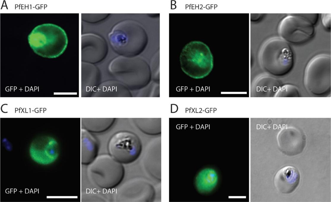
Four a/b-hydrolases are exported into the erythrocyte compartment. Immunofluorescence assay (IFA) of trophozoite/early schizont-stage parasites overexpressing GFP-tagged a/b-hydrolases. Parasites were fixed with paraformaldehyde/glutaraldehyde and stained with anti-GFP (green). The nuclei were stained with 4’,6’-iamidino-2-phenylindole (DAPI) (blue). (A) PfEH1-GFP, (B) PfEH2-GFP, (C) PfXL1-GFP, and (D) PfXL2-GFP. Expression was under control of the constitutive HSP86 promoter. The images are representative of at least three independent experiments. Bars=5 mm. DIC, differential interference contrast. For the two a/b-hydrolase familyA members, fluorescence was observed at the periphery of the infected erythrocyte and within the parasite (A and B). In contrast, for the two family B members, fluorescence was diffuse throughout the cytosol of the infected RBC (C and D).Spillman NJ, Dalmia VK, Goldberg DE. Exported Epoxide Hydrolases Modulate Erythrocyte Vasoactive Lipids during Plasmodium falciparum Infection. MBio. 2016 Oct 18;7(5). pii: e01538-16.
See original on MMPMore information
| PlasmoDB | PF3D7_0301300 |
| GeneDB | PF3D7_0301300 |
| Malaria Metabolic Pathways | Localisation images Pathways mapped to |
| Previous ID(s) | PFC0065c |
| Orthologs | |
| Google Scholar | Search for all mentions of this gene |