PF3D7_0207500 serine repeat antigen 6 (SERA6)
Disruptability [+]
| Species | Disruptability | Reference | Submitter |
|---|---|---|---|
| P. falciparum 3D7 |
Refractory |
22984267 | Theo Sanderson, Wellcome Trust Sanger Institute |
| P. falciparum 3D7 |
Refractory |
USF piggyBac screen (Insert. mut.) | USF PiggyBac Screen |
Mutant phenotypes [+]
| Species | Stage | Phenotype | Reference | Submitter |
|---|---|---|---|---|
| P. falciparum 3D7 | Asexual |
Egress defect |
29459732 DiCre approach showed essentiality of SERA6. "PVM rupture and RBC membrane poration occur normally in SERA6-null parasites but RBC membrane rupture does not occur." |
Theo Sanderson, Wellcome Trust Sanger Institute |
Imaging data (from Malaria Metabolic Pathways)
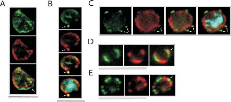
Colocalization of ETRAMPs with EXP-1 and SERP. The first image in each set shows ETRAMP labeling (green, FITC), the second Texas-Red-labeled SERP (A) or EXP-1 (B–F), and the third image represents the merged picture. The fourth panel in B and C depicts the merged picture, including nuclear staining (DAPI). (A) Confocal microscopy analysis of SERP and ETRAMP4 colocalization in schizont stage parasites (dried and 1% formaldehyde fixed). The ETRAMP4 signal seems partially detached from the PV content (arrow). (B and C) ETRAMP4 and EXP-1 localize to dots, probably representing vesicular structures that are detached from the PVM (E) or seem connected to the PVM (F). Dots are indicated by arrows. (D and E) Fluorescent microscopic analysis of ETRAMP and EXP-1 costaining of a ring-stage parasite displaying an even circular pattern (D, 0.1% formaldehyde-fixed IRBCs, ETRAMP10.1) or a beads on a string pattern (E, unfixed IRBCs, ETRAMP2). Yellow color shows regions of overlap (arrows). This indicated that ETRAMPs and EXP-1 localize to different regions of the PVM.Spielmann T, Fergusen DJ, Beck HP. etramps, a new Plasmodium falciparum gene family coding for developmentally regulated and highly charged membrane proteins located at the parasite-host cell interface. Mol Biol Cell. 2003 14:1529-44.
See original on MMP
Immunoelectron micrograph of a P. falciparum schizont section reacted with an antiserum raised against the fusion protein expressed by pEX41-10b. x 18 000. pEX140-10b codes for 21 kDa polypeptide resulting from the fusion of the SERA6 cDNA and the lgt11 plasmid. The SERA6 antigen was found in the soluble as well as in the membrane fraction of schizonts, but was not detected in isolated merozoites by Western blot analysis. Immunoelectron microscopy using gold-labeled second antibody showed the antigen to be localized mainly within the parasitophorous vacuole.Knapp B, Nau U, Hundt E, Küpper HA. A new blood stage antigen of Plasmodium falciparum highly homologous to the serine-stretch protein SERP. Mol Biochem Parasitol. 1991 44:1-13. Copyright Elsevier 2010
See original on MMP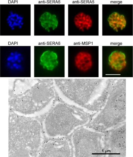
SERA6 is a soluble parasitophorous vacuole (PV) protein which may partially interact with the PVM. Upper panel: IFA demonstrates colocalization of the anti-SERA6 and anti-SERA5 signal in mature schizonts, except that the SERA6 signal additionally shows an association with the outer confines of the intracellular parasite, probably corresponding to the PVM. Scale bar, 5 μm. Lower panel: Immuno-electron microscopic localization of SERA6 in a P. falciparum schizont, using the anti-S6C1 antibodies labelled with 10 nm immunogold. The majority of the signal is clearly associated with the peripheral space between intracellular merozoites, consistent with a PV localization. Magnification, x 15000.Ruecker A, Shea M, Hackett F, Suarez C, Hirst EM, Milutinovic K, Withers-Martinez C, Blackman MJ. Proteolytic activation of the essential parasitophorous vacuole cysteine protease SERA6 accompanies malaria parasite egress from its host erythrocyte. J Biol Chem. 2012287(45):37949-63.
See original on MMP
SERA3–6 genes are expressed in single parasites at similar localization. Trophozoite- and schizont (Honduras-1)-infected erythrocytes were purified by Percoll and subjected to immunofluorescence staining with rabbit anti-SE3N, -SE4N, or -SE6N antiserum and mouse anti-SE47’ (SERA5) IgG. The secondary antibodies used were Cy3-conjugated anti-rabbit IgG and fluorescein isothiocyanate-conjugated anti-mouse IgG. 4’,6’-Diamidino-2-phenylindole was also used to stain parasite nuclei.Aoki S, Li J, Itagaki S, Okech BA, Egwang TG, Matsuoka H, Palacpac NM, Mitamura T, Horii T. Serine repeat antigen (SERA5) is predominantly expressed among the SERA multigene family of Plasmodium falciparum, and the acquired antibody titers correlate with serum inhibition of the parasite growth. J Biol Chem. 2002 277:47533-40.
See original on MMPMore information
| PlasmoDB | PF3D7_0207500 |
| GeneDB | PF3D7_0207500 |
| Malaria Metabolic Pathways | Localisation images Pathways mapped to |
| Previous ID(s) | PF02_0071, PFB0335c |
| Orthologs | PBANKA_0304800 , PBANKA_0304900 , PBANKA_0305000 , PBANKA_0305100 , PCHAS_0307000 , PCHAS_0307100 , PCHAS_0307200 , PCHAS_0307300 , PKNH_0413100 , PKNH_0413200 , PKNH_0413400 , PKNH_0413600 , PKNH_0413700 , PVP01_0416800 , PVP01_0416900 , PVP01_0417000 , PVP01_0417100 , PVP01_0417200 , PVP01_0417300 , PVP01_0417400 , PVP01_0417600 , PVP01_0417700 , PVP01_0417800 , PVP01_0417900 , PVX_003795 , PVX_003800 , PVX_003805 , PVX_003810 , PVX_003820 , PVX_003825 , PVX_003830 , PVX_003835 , PVX_003840 , PVX_003845 , PVX_003850 , PY17X_0305600 , PY17X_0305700 |
| Google Scholar | Search for all mentions of this gene |