PF3D7_0206800 merozoite surface protein 2 (MSP2)
Disruptability [+]
| Species | Disruptability | Reference | Submitter |
|---|---|---|---|
| P. falciparum 3D7 |
Refractory |
22986493 | Theo Sanderson, Wellcome Trust Sanger Institute |
| P. falciparum 3D7 |
Possible |
USF piggyBac screen (Insert. mut.) | USF PiggyBac Screen |
Mutant phenotypes [+]
None reported yet. Please press the '+' button above to add one.Imaging data (from Malaria Metabolic Pathways)
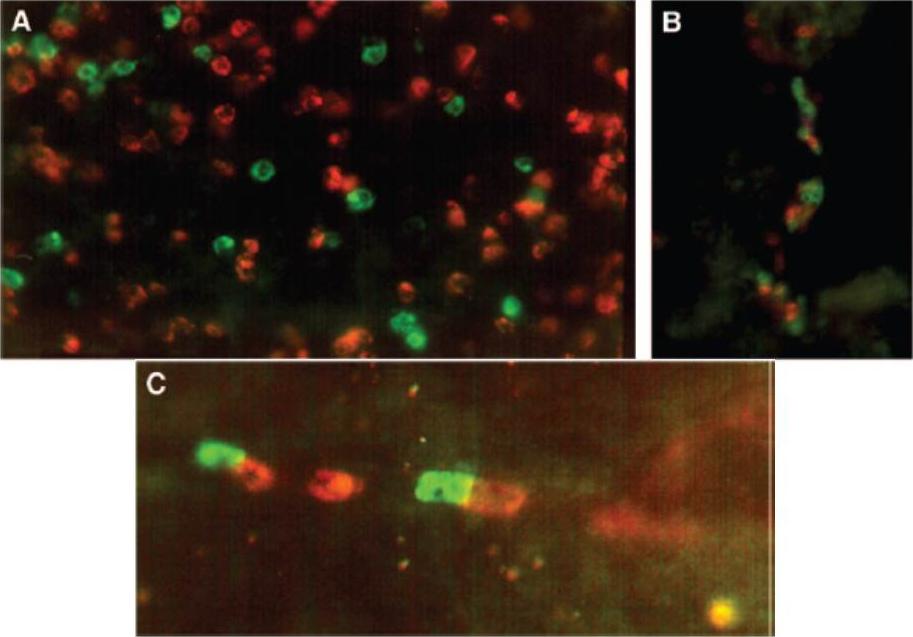
Detection of multiple-clone infections in blood and brain by double-labeled IFAT: MSP-1 serotypes of P. falciparum in peripheral blood and brain tissue of patient 13. (A and B) P. falciparum schizont-infected erythrocytes cultured from the peripheral blood (A) and homogenate from the frontal lobe of the brain (B) of the same child. A majority of schizonts (red) reacted with MSP-1 block 4-specific MAb 10-2B (immunoglobulin G2a) plus RITC-conjugated anti-immunoglobulin G2a. A minority of schizonts (green) were positive with MAb 12.1 (immunoglobulin G1) plus FITC-conjugated anti-immunoglobulin G1. The detectable serotypes or relative proportions of sequestered and circulating parasites did not differ. Magnification, 630. (C) Thin smear prepared from a supraorbital needle sample from patient SO, showing a mixed infection of two MSP-2 serotypes in a brain capillary. Some schizonts (red) reacted with MSP-2 group B-specific MAb 8G10/48 (immunoglobulin G2b) plus RITC conjugated anti-immunoglobulin G2b, whereas others (green) reacted with MSP-2 group A-specific MAb 12.3 (immunoglobulin G1) plus FITC-conjugated anti-immunoglobulin G1. Magnification, 630.Dobaño C, Rogerson SJ, Taylor TE, McBride JS, Molyneux ME. Expression of merozoite surface protein markers by Plasmodium falciparum-infected erythrocytes in peripheral blood and tissues of children with fatal malaria. Infect Immun. 2007 75:643-52.
See original on MMP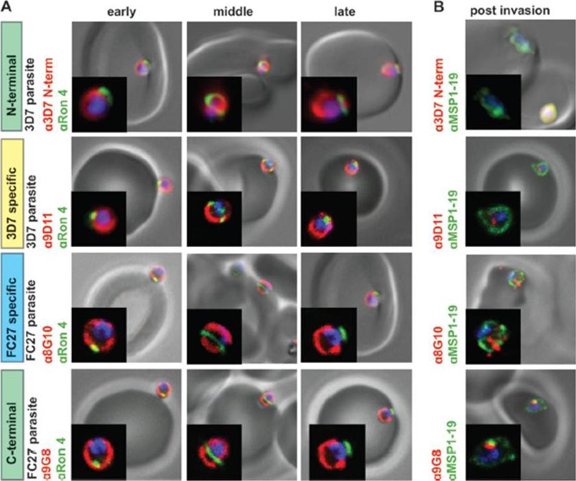
GPI-anchored MSP2 is carried into invaded RBCs and rapidly degraded. (A) Invading merozoites were labeled with the tight junction marker PfRON4(green) and colabeled with antibodies to different regions of MSP2 (red). All regions of MSP2 were carried through the tight junction into the RBC, with labeling being visible on both sides of PfRON4 at the tight junction. (B) Newly invaded rings (10 min postinvasion) were labeled with antibodies to MSP1-19 (green) and antibodies to different regions of MSP2 (red). MSP2 was rapidly degraded postinvasion, with little or no labeling being visible in rings. Images are single slices from deconvoluted stacks. Representative images of antibodies to the different regions of MSP2; other antibodies tested showed the same labeling patterns. The MSP2 allele of the parasite strain used in each row of images is indicated as 3D7 parasite or FC27 parasite (D10 parasite strain). Antibody dilutions used were 1:500 for secondary antibodies, 1:250 for 8G10, 1:100 for PfRON4, and 1:50 for N-terminal purified antibodies, 9D11, and9G8.Boyle MJ, Langer C, Chan JA, Hodder AN, Coppel RL, Anders RF, Beeson JG. Sequential processing of merozoite surface proteins during and after erythrocyte invasion by Plasmodium falciparum. Infect Immun. 2014 82(3):924-36.
See original on MMP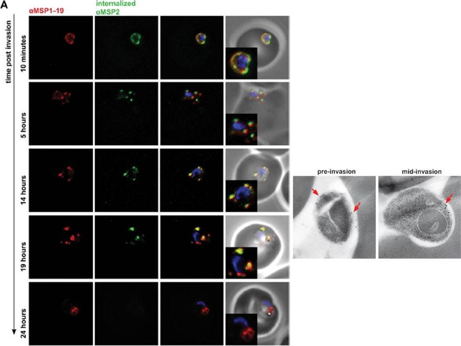
Parasites were labeled with polyclonal rabbit antibodies to MSP1-19 and secondary antibodies (anti-rabbit Alexa594 to detect MSP1-19 labelingand anti-mouse Alexa488 to detect internalized MSP2 MAbs, with anti-MSP1-19 at a 1:200 dilution and secondary antibodies at a 1:500 dilution).Internalized MSP2 MAbs were detected up to 19 h postinvasion. Images are from representative antibodies to the C-terminally conserved region. The labeling of internalized MSP2 MAb was colocalizedwith labeling of MSP1-19 by polyclonal rabbit antibodies, further suggesting that MAbs to MSP2 were bound to the parasite surface postinvasion. Lower panel: 3D7 strain merozoites were fixed during invasion for EM and labeled with MSP2 N-terminal antibodies. A repre-sentative image of a merozoite prior to and during invasion is shown. Labeling is indicated with red arrows.Boyle MJ, Langer C, Chan JA, Hodder AN, Coppel RL, Anders RF, Beeson JG. Sequential processing of merozoite surface proteins during and after erythrocyte invasion by Plasmodium falciparum. Infect Immun. 2014 82(3):924-36.
See original on MMPMore information
| PlasmoDB | PF3D7_0206800 |
| GeneDB | PF3D7_0206800 |
| Malaria Metabolic Pathways | Localisation images Pathways mapped to |
| Previous ID(s) | PF02_0064, PFB0300c |
| Orthologs | |
| Google Scholar | Search for all mentions of this gene |