PF3D7_0202400 gamete antigen 27/25, putative
Disruptability [+]
| Species | Disruptability | Reference | Submitter |
|---|---|---|---|
| P. falciparum 3D7 |
Possible |
28288121 SLI |
Theo Sanderson, Wellcome Trust Sanger Institute |
| P. falciparum 3D7 |
Possible |
USF piggyBac screen (Insert. mut.) | USF PiggyBac Screen |
Mutant phenotypes [+]
| Species | Stage | Phenotype | Reference | Submitter |
|---|---|---|---|---|
| P. falciparum 3D7 | Asexual |
No difference |
28288121 SLI |
Theo Sanderson, Wellcome Trust Sanger Institute |
Imaging data (from Malaria Metabolic Pathways)
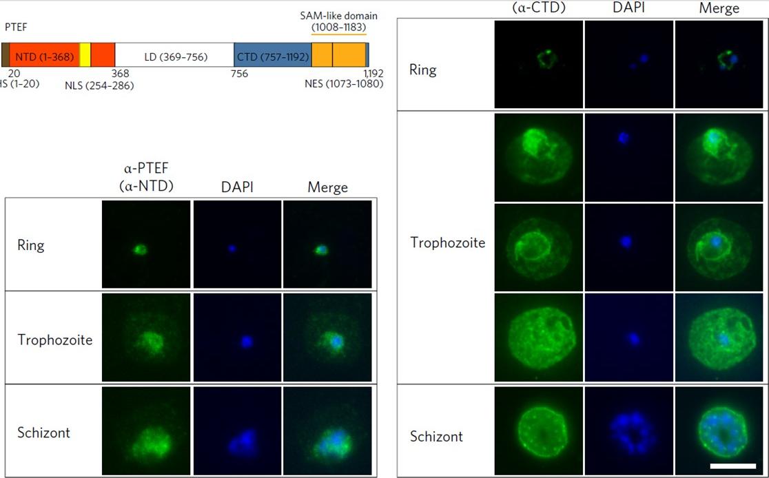
PTEF localizes to multiple cellular compartments. Immunofluorescence assay (IFA) using anti-NTD serum (left) and anti-CTD IgG (right). DAPI was used to stain parasite DNA. Images are representative of five IFA replicates. Scale bars, 5 μm. During ring stages, CTD staining was localized to the cytoplasm of the parasite, whereas late-stage parasites displayed strong fluorescence signals that covered both the parasitic and erythrocytic compartmentsin most IEs, resembling the pattern of full-length GFP fusion transfectants in the live imaging. However, heterogeneous staining was observed for some late-stage IEs, with some showing predominant localization within the parasite compartment, whereas others exhibited preferential staining at the PV/PPM. Despite the lack of a PEXEL motif, the export of the PTEF protein was also observed when using antibodies against the NTD, although a lower erythrocytic-to-parasitic signal ratiowas observed.Chan S, Frasch A, Mandava CS, Ch'ng JH, Quintana MDP, Vesterlund M, Ghorbal M, Joannin N, Franzén O, Lopez-Rubio JJ, Barbieri S, Lanzavecchia A, Sanyal S, Wahlgren M. Regulation of PfEMP1-VAR2CSA translation by a Plasmodium translation-enhancing factor. Nat Microbiol. 2017 May 8;2:17068.
See original on MMP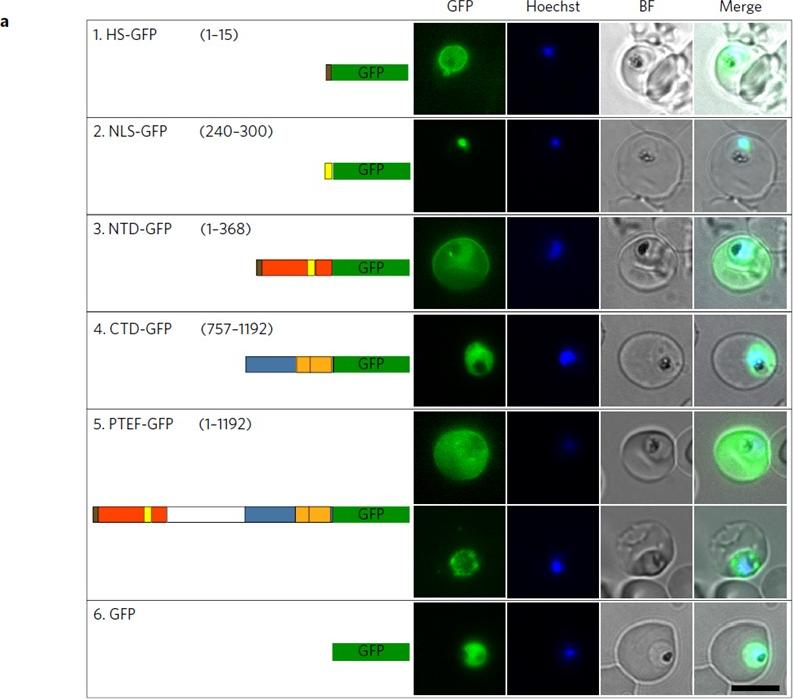
PTEF localizes to multiple cellular compartments. a, Direct live imaging on parasites transfected with GFP genes fused with HS (1), NLS (2), NTD (3), CTD (4), full-length PTEF (5) and GFP alone (6). HS-GFP displayed exclusive localization to the parasitophorous vacuole (PV)/parasite plasma membrane (PPM), whereas the NLS-GFP construct faithfully imported GFP into the nucleus. Interestingly, the NLS signal was overridden when present in the whole NTD, resulting in diffuse localization of the NTD-GFP to the PV/PPM as well asto the erythrocyte cytoplasm and membrane. By contrast, CTD-GFP was only found to localize in the parasite cytoplasm. Again, the full-length PTEF-GFP displayed a diffuse pattern in the entire parasite and host erythrocytes, with a small fraction of IEs showing intense localization to the PV/PPM.Chan S, Frasch A, Mandava CS, Ch'ng JH, Quintana MDP, Vesterlund M, Ghorbal M, Joannin N, Franzén O, Lopez-Rubio JJ, Barbieri S, Lanzavecchia A, Sanyal S, Wahlgren M. Regulation of PfEMP1-VAR2CSA translation by a Plasmodium translation-enhancing factor. Nat Microbiol. 2017 May 8;2:17068.
See original on MMPMore information
| PlasmoDB | PF3D7_0202400 |
| GeneDB | PF3D7_0202400 |
| Malaria Metabolic Pathways | Localisation images Pathways mapped to |
| Previous ID(s) | PF02_0024, PFB0115w |
| Orthologs | |
| Google Scholar | Search for all mentions of this gene |