PF3D7_0202200 EMP1-trafficking protein (PTP1)
Mutant phenotypes [+]
None reported yet. Please press the '+' button above to add one.Imaging data (from Malaria Metabolic Pathways)
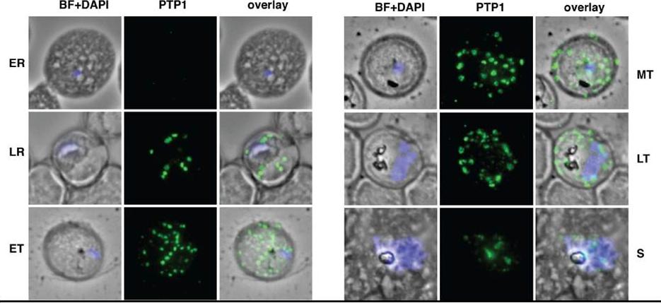
Localisation of PfPTP1 during the intracellular cycle in immunofluorescence assay. The fixed cells were incubated with α-PTP1 antibody. Parasite nucleus was stained with DAPI. ER = early ring, LR=late ring, ET=early trophozoite, MT=mid trophozoite, LT=late trophozoite and S=rupturing schizont. BF = bright field. PTP1 was expressed across the asexual cycle and located in punctate structures, surrounding the intracellular parasite, that bud from the PVM. During development these structures increase in size contacting the RBC membrane in the mature parasite . In rupturing schizonts PfPTP1 associated organelles appear as remnants between merozoites.Rug M, Cyrklaff M, Mikkonen A, Lemgruber L, Kuelzer S, Sanchez CP, Thompson J, Hanssen E, O'Neill M, Langer C, Lanzer M, Frischknecht F, Maier AG, Cowman AF. Export of virulence proteins by malaria-infected erythrocytes involves remodelling of host actin cytoskeleton. Blood. 2014 Aug 19 [Epub ahead of print]
See original on MMP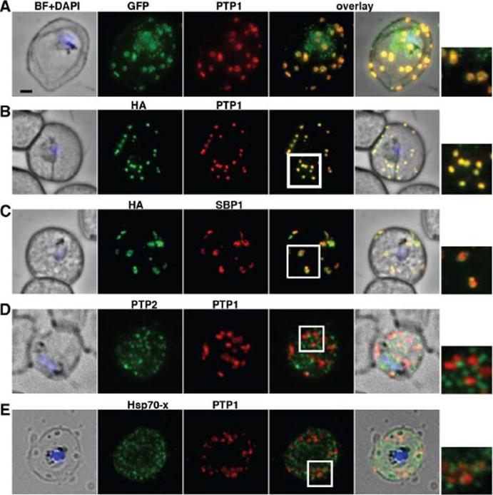
PfPTP1 is localised on Maurer’s clefts in P. falciparum-infected RBCs. (A) Immunofluorescence assay on CS2/PfPTP1-GFP cell line: the GFP signal (green) colocalises with that of the endogenous protein (PfPTP1; red); inset: enlargement of colocalisation pattern. (B) Immuno-fluorescence assay on CS2/PfPTP1-HA cell line: the HA signal (green) colocalises with the α-PfPTP1 signal (red); inset: enlargement of colocalisation pattern. (C) Immunofluorescence assay on CS2/PfPTP1-HA cell line: the HA-signal (green) colocalises partially with the MC resident protein SBP1 (red); inset: enlargement of colocalisation pattern. (D) Immunofluorescence assay on CS2 parental cell line: the PfPTP2-signal (green) does not overlap with the PfPTP1-signal (red); inset: enlargement of area where MC (PfPTP1) and electron dense vesicles (PfPTP2) are in close proximity. (E) Immunofluorescence assay on CS2 parental cell line: the PfHsp70-x-signal (green) colocalises partially with the PfPTP1- signal (red); inset: enlargement of partial colocalisation on J-Dots (PfHsp70-x). Scale bar: 1μm; same size of ROI in each image. Rug M, Cyrklaff M, Mikkonen A, Lemgruber L, Kuelzer S, Sanchez CP, Thompson J, Hanssen E, O'Neill M, Langer C, Lanzer M, Frischknecht F, Maier AG, Cowman AF. Export of virulence proteins by malaria-infected erythrocytes involves remodelling of host actin cytoskeleton. Blood. 2014 Aug 19 [Epub ahead of print]
See original on MMP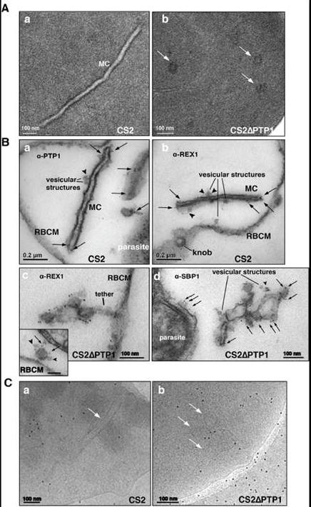
Comparison of transmission electron microscopical studies on CS2 versus CS2ΔPTP1 cell line.(A) Conventional chemical Fixation: a) CS2 parental line. MC (MC) display electron dense membranes and an electron lucent lumen; b) CS2ΔPTP1 cell line shows globular structures and no long lamellar MC structures (arrows). (B) Equinatoxin II treated cells with pre-embedding labelling: a) α-PfPTP1 antibodies or b) α-REX1 antibodies label the slender MC membranes (arrows) and vesicles (arrowheads) in the CS2 parasite line; c) α-REX1 or d) α-SBP1 antibodies label globular structures (arrows) and vesicular structures (arrowheads) in the CS2ΔPTP1 cell line. RBCM – red blood cell membrane. (C) The frozen hydrated samples show MC (arrow) in which the lumen looks similar to the RBC cytoplasm as described earlier. The CS2ΔPTP1 cell line shows aggregated globular structures (arrows). In CS2 typical single lamellae for MC were observed with an electron dense coat and translucent lumen (Aa). In contrast, CS2ΔPTP1 displayed electron dense areas randomly distributed through the RBC cytoplasm (Ab). The α-PfPTP1 antibodies line the MC lamella similar to REX1 localisation (B). Vesicular structures were observed that appeared to bud off MC and these had a dense lumen and less distinct membranes. These may be vesicles involved in trafficking between PVM and MC and/or MC to the RBC membrane (i.e. J-Dots). These structures were decorated withPfPTP1 and REX1 antibodies (Ba,b). In CS2ΔPTP1 the architecture of MC was disrupted showing large aggregated globular structures (Bc,d). SBP1 and REX1 were located on these structures. Single lamellae of MC were observed in CS2 (Ca) whereas globular structures were observed for CS2ΔPTP1 (Cb).Rug M, Cyrklaff M, Mikkonen A, Lemgruber L, Kuelzer S, Sanchez CP, Thompson J, Hanssen E, O'Neill M, Langer C, Lanzer M, Frischknecht F, Maier AG, Cowman AF. Export of virulence proteins by malaria-infected erythrocytes involves remodelling of host actin cytoskeleton. Blood. 2014 Aug 19 [Epub ahead of print]
See original on MMPMore information
| PlasmoDB | PF3D7_0202200 |
| GeneDB | PF3D7_0202200 |
| Malaria Metabolic Pathways | Localisation images Pathways mapped to |
| Previous ID(s) | PF02_0022, PFB0106c |
| Orthologs | |
| Google Scholar | Search for all mentions of this gene |