PF3D7_0201800 knob associated heat shock protein 40 (KAHsp40)
Disruptability [+]
| Species | Disruptability | Reference | Submitter |
|---|---|---|---|
| P. falciparum 3D7 |
Possible |
18614010 | Theo Sanderson, Wellcome Trust Sanger Institute |
| P. falciparum 3D7 |
Possible |
USF piggyBac screen (Insert. mut.) | USF PiggyBac Screen |
Mutant phenotypes [+]
None reported yet. Please press the '+' button above to add one.Imaging data (from Malaria Metabolic Pathways)
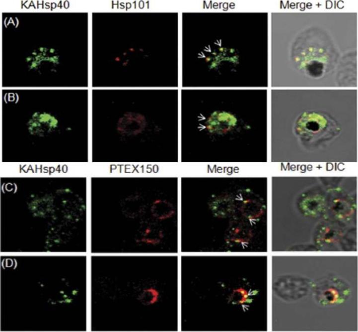
KAHsp40 associates with the PEXEL translocon on the PVM. (A, B) IFA analysis reveals that KAHsp40 and Hsp101 co-localize with each other on the PVM. (C, D) IFA analysis reveals that KAHsp40 and PTEX150 co-localize with each other on the PVM. White arrows indicate the discrete foci in which they co-localize.Acharya P, Chaubey S, Grover M, Tatu U. An Exported Heat Shock Protein 40 Associates with Pathogenesis-Related Knobs in Plasmodium falciparum Infected Erythrocytes. PLoS One. 2012;7(9):e44605.
See original on MMP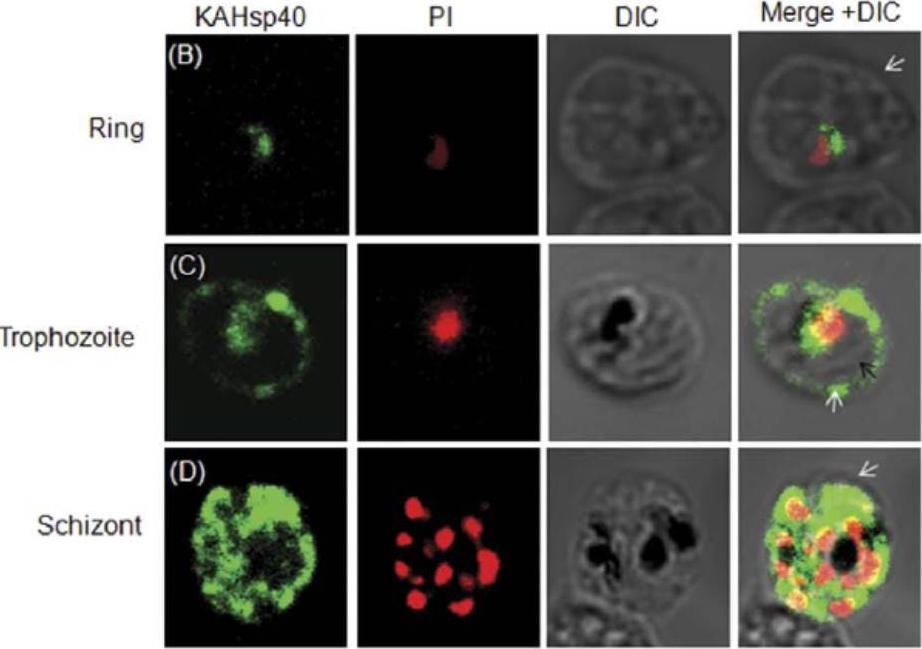
KAHsp40 is exported into the infected RBC cytosol. IFA of (B) ring stage parasites (C) trophozoite stage parasites (D) schizont stage parasites reveals the presence of KAHsp40 in discrete foci in the erythrocyte compartment. Black arrow indicates the parasite boundary and white arrow indicates the erythrocyte membrane.Acharya P, Chaubey S, Grover M, Tatu U. An Exported Heat Shock Protein 40 Associates with Pathogenesis-Related Knobs in Plasmodium falciparum Infected Erythrocytes. PLoS One. 2012;7(9):e44605.
See original on MMP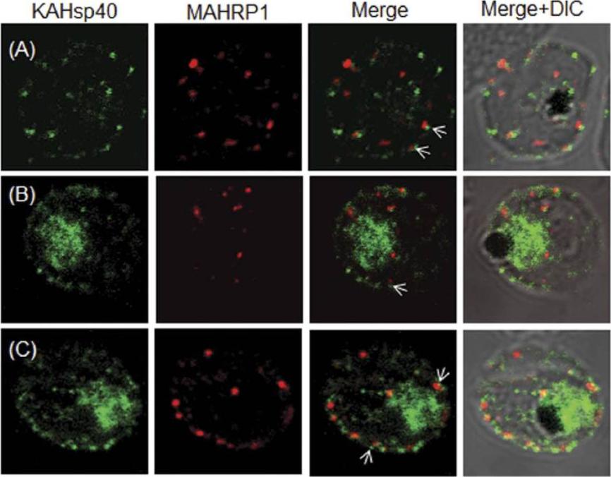
KAHsp40 does not associate with Maurer’s cleft. (A–C) IFA reveals that both KAHsp40 and MAHRP1 are present in discrete and different foci in the infected erythrocyte, however, they do not co-localize with each other in spite of signals being in close proximity (highlighted by white arrows). The images shown have been taken at the trophozoite stage.Acharya P, Chaubey S, Grover M, Tatu U. An Exported Heat Shock Protein 40 Associates with Pathogenesis-Related Knobs in Plasmodium falciparum Infected Erythrocytes. PLoS One. 2012;7(9):e44605.
See original on MMP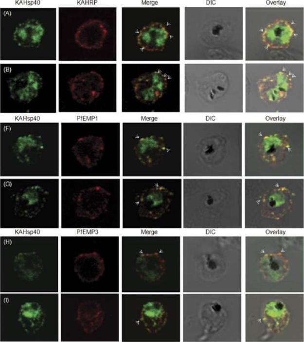
KAHsp40 associates with knobs on the infected erythrocyte membrane. (A-B) IFA reveals that KAHsp40 and KAHRP co-localize in the erythrocyte periphery near the erythrocyte plasma membrane. White arrows indicate the discrete foci in which they co-localize. KAHsp40 and PfEMP1 and KAHsp40 and PfEMP3 co-localize with each other on the erythrocyte plasma membrane indicating the close association of KAHsp40 with knobs.Acharya P, Chaubey S, Grover M, Tatu U. An Exported Heat Shock Protein 40 Associates with Pathogenesis-Related Knobs in Plasmodium falciparum Infected Erythrocytes. PLoS One. 2012;7(9):e44605.
See original on MMP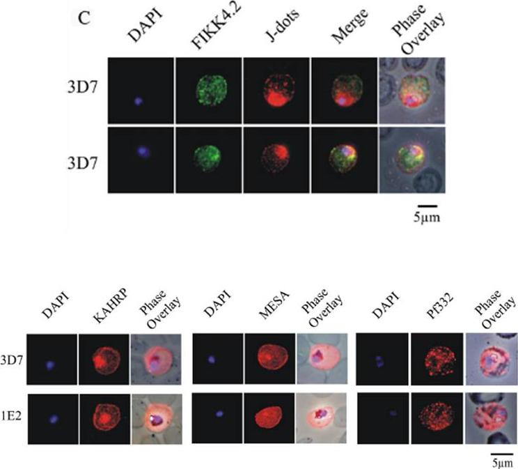
(C) Immunofluorescence analysis of red blood cells infected with parental 3D7 parasites co-labelled with a-FIKK4.2 and a-PFB0090c (J-dots) antibodies. Merge and phase overlay images indicate that FIKK4.2 and J-dots do not co-localise in infected red blood cells. In all panels, parasite nuclei are counterstained with DAPI. (D) Immunofluorescence analysis of red blood cells infected with either parental 3D7 or FIKK4.2 knockout (1E2) parasites. Infected red blood cells were incubated with antibodies raised against the knob-associated histidine-rich protein (KAHRP), mature-parasite-infected erythrocyte surface antigen (MESA) or P. falciparum antigen 332 (Pf332). Parasite nuclei were counterstained with DAPI. KAHRP, MESA and Pf332 as three examples of other very well described P. falciparum-encoded exported proteins.Kats LM, Fernandez KM, Glenister FK, Herrmann S, Buckingham DW, Siddiqui G, Sharma L, Bamert R, Lucet I, Guillotte M, Mercereau-Puijalon O, Cooke BM. An exported kinase (FIKK4.2) that mediates virulence-associated changes in Plasmodium falciparum-infected red blood cells. Int J Parasitol. 2014 Feb 14. [Epub ahead of print]
See original on MMP
Localization of LyMP in iRBCs. A) Localization of LyMP in trophozoites, with either anti-LyMP (green) or anti-HA (red) showing that LyMP is exported into the RBC cytosol. 4’,6’-Diamidino-2-phenylidole (DAPI) is a nuclear stain (blue). B) Colocalization of LyMP (green) with a variety of iRBC markers (red) that are indicated in the panel. DAPI is a nuclear stain (blue). C) High-resolution structured illumination fluorescence microscopy (OMX) with anti-LyMP (green) and either anti-KAHRP, anti-MESA, or anti-spectrin (red) colocalizations. Corresponding overlay images are shown for all. D) Immunoelectron micrograph of gold-labeled Ha-tagged parasites. Red asterisks indicate knobs and the arrow indicates gold labeling of LyMP at the RBC membrane skeleton. P, parasite; MC, Maurer’s clefts. LyMP showed a pattern of distribution similar to that for MESA, KAHRP. LyMP did not consistently colocalize with MCs or to other punctate structures previously identified in the iRBC cytosol such as those seen for FIKK4.2 (R45) or for J-dots (KAHsp40).Proellocks NI, Herrmann S, Buckingham DW, Hanssen E, Hodges EK, Elsworth B, Morahan BJ, Coppel RL, Cooke BM. A lysine-rich membrane-associated PHISTb protein involved in alteration of the cytoadhesive properties of Plasmodium falciparum-infected red blood cells. FASEB J. 2014 Apr 4. [Epub ahead of print]
See original on MMPMore information
| PlasmoDB | PF3D7_0201800 |
| GeneDB | PF3D7_0201800 |
| Malaria Metabolic Pathways | Localisation images Pathways mapped to |
| Previous ID(s) | PF02_0018, PFB0090c |
| Orthologs | |
| Google Scholar | Search for all mentions of this gene |