PF3D7_0201600 PHISTb domain-containing RESA-like protein 1 (RLP1)
Disruptability [+]
| Species | Disruptability | Reference | Submitter |
|---|---|---|---|
| P. falciparum 3D7 |
Possible |
USF piggyBac screen (Insert. mut.) | USF PiggyBac Screen |
| P. falciparum 3D7 |
Possible |
25342752 \"Disruption of PFB0080c resulted in increased var2csa transcription and VAR2CSA surface expression, leading to higher C4S-binding capacity of infected erythrocytes. Further, PFB0080c-knock-out parasites stably maintained the C4S adherence through many generations of growth. Although the majority of PFB0080c-knock-out parasites bound to C4S even after culturing for 6 months, a minor population bound to both C4S and CD36. These results strongly suggest that the loss of PFB0080c markedly compromises the var gene switching process, leading to a marked reduction in the switching rate and additional PfEMP1 expression by a minor population of parasites\" |
Theo Sanderson, Francis Crick Institute |
Mutant phenotypes [+]
| Species | Stage | Phenotype | Reference | Submitter |
|---|---|---|---|---|
| P. falciparum 3D7 | Asexual |
Transcriptional effect |
25342752 \"Disruption of PFB0080c resulted in increased var2csa transcription and VAR2CSA surface expression, leading to higher C4S-binding capacity of infected erythrocytes. Further, PFB0080c-knock-out parasites stably maintained the C4S adherence through many generations of growth. Although the majority of PFB0080c-knock-out parasites bound to C4S even after culturing for 6 months, a minor population bound to both C4S and CD36. These results strongly suggest that the loss of PFB0080c markedly compromises the var gene switching process, leading to a marked reduction in the switching rate and additional PfEMP1 expression by a minor population of parasites\" |
Theo Sanderson, Francis Crick Institute |
Imaging data (from Malaria Metabolic Pathways)

Localisation of PHIST:GFP proteins. The left- and right-hand images show GFP localisation and a phase contrast image, respectively. White filled arrows: peripheral GFP puncta; White unfilled arrow: GFP puncta in host erythocyte cytosol. Scale bar, 2 mm. GFP-tagged PF3D7_0401800, PF3D7_0424600, PF3D7_0532400, PF3D7_1102500 and PF3D7_1476200 were all exported to the host cell and displayed a striking localisation at the edge of the host erythrocyte (1A–E), PF3D7 1252700 was clearly peripheral in the host erythrocyte (F). PF3D7_0201600 (G) shows weak accumulation at the erythrocyte periphery, but the majority of the protein was localised in the RBC cytosol. PF3D7_0936600 was localised in the erythrocyte cytosol, not at the host cell periphery (H).Tarr SJ, Moon RW, Hardege I, Osborne AR. A conserved domain targets exported PHISTb family proteins to the periphery of Plasmodium infected erythrocytes. Mol Biochem Parasitol. 2014 196(1):29-40 PMID: 25106850
See original on MMP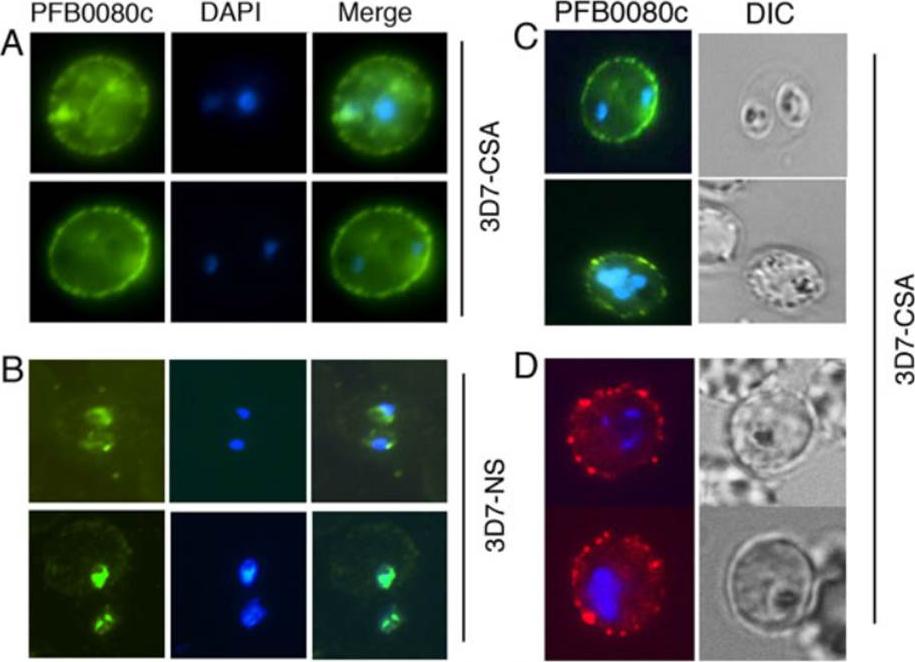
PFB0080c is expressed on the IRBC surface of C4S-selected P. falciparum. A-D, Immunofluorescence analysis of IRBCs was performed using anti-PFB0080c antiserum followed by either FITC-conjugated goat anti-mouse IgG (A-C) or phycoerythrin-conjugated goat antimouse IgG (D) secondary antibody. A and B, Permeabilized C4S-adherent (3D7-CSA) IRBCs (A) and non-selected (3D7-NS) IRBCs (B). C and D, Live 3D7-CSA IRBCs. PFB0080c is strongly expressed on the surface of C4S adherent IRBCs, but not in 3D7-NS IRBCs.Goel S, Muthusamy A, Miao J, Cui L, Salanti A, Winzeler EA, Gowda DC. Targeted disruption of a ring-infected erythrocyte surface antigen (RESA)-like export protein gene in Plasmodium falciparum confers stable chondroitin 4-sulfate cytodherence capacity. J Biol Chem. 2014 Oct 23. [Epub ahead of print]
See original on MMP
PFB0080c-knockout P. falciparum express VAR2CSA on the surface of infected erythrocytes. A and B, Immuno-fluorescence analysis of VAR2CSA in live ΔPFB0080c IRBCs was performed using rabbit anti-VAR2CSA antiserum followed by biotin-conjugated sheep antirabbit IgG secondary antibody and phycoerythrin-conjugated streptavidin. Shown are the immunofluorescent and light micrographs of parental 3D7-CSA parasites (A) and ΔPFB0080c parasites (B); both parasite types expressed VAR2CSA protein. Goel S, Muthusamy A, Miao J, Cui L, Salanti A, Winzeler EA, Gowda DC. Targeted disruption of a ring-infected erythrocyte surface antigen (RESA)-like export protein gene in Plasmodium falciparum confers stable chondroitin 4-sulfate cytodherence capacity. J Biol Chem. 2014 Oct 23. [Epub ahead of print]
See original on MMP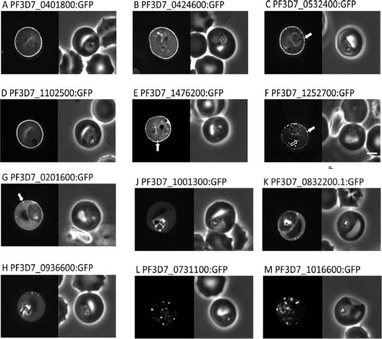
Localisation of PHIST:GFP proteins. The left- and right-hand images show GFP localisation and a phase contrast image, respectively. White filled arrows: peripheral GFP puncta; White unfilled arrow: GFP puncta in host erythocyte cytosol. Scale bar, 2 mm. GFP-tagged PF3D7_0401800, PF3D7_0424600, PF3D7_0532400, PF3D7_1102500 and PF3D7_1476200 were all exported to the host cell and displayed a striking localisation at the edge of the host erythrocyte (1A–E), PF3D7 1252700 was clearly peripheral in the host erythrocyte (F). PF3D7_0201600 (G) shows weak accumulation at the erythrocyte periphery, but the majority of the protein was localised in the RBC cytosol. PF3D7_0936600 was localised in the erythrocyte cytosol, not at the host cell periphery (H).Tarr SJ, Moon RW, Hardege I, Osborne AR. A conserved domain targets exported PHISTb family proteins to the periphery of Plasmodium infected erythrocytes. Mol Biochem Parasitol. 2014 196(1):29-40
See original on MMPMore information
| PlasmoDB | PF3D7_0201600 |
| GeneDB | PF3D7_0201600 |
| Malaria Metabolic Pathways | Localisation images Pathways mapped to |
| Previous ID(s) | PF02_0016, PFB0080c |
| Orthologs | |
| Google Scholar | Search for all mentions of this gene |