PF3D7_0101300 Pfmc-2TM Maurer's cleft two transmembrane protein (MC-2TM)
Disruptability [+]
| Species | Disruptability | Reference | Submitter |
|---|---|---|---|
| P. falciparum 3D7 |
Possible |
USF piggyBac screen (Insert. mut.) | USF PiggyBac Screen |
Mutant phenotypes [+]
None reported yet. Please press the '+' button above to add one.Imaging data (from Malaria Metabolic Pathways)
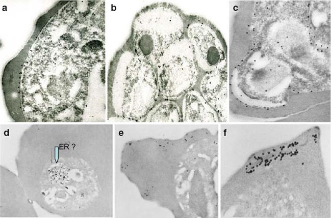
Immunoelectron micrographs of young trophozoite-, late trophozoite-, young schizont-, and late schizont-infected erythrocytes reactive with rabbit antisera against PfMC-2TM peptides, 703, 705, and 713 (identical sequences in all PfMC-2TM-coding genes and Mab SP1A6 against the 130 kDa Maurer’s cleft protein. a–c Peptide-specific antisera are reactive with the PV/PVM and weakly around clefts. d–f Mab SP1A6 is reactive with structures within the parasite in young trophozoites, probably the endoplasmic reticulum (d), late trophozoites in erythrocyte cytoplasm under knobs (e), and in young schizonts around longitudinal clefts in the erythrocyte cytoplasm (f).Tsarukyanova I, Drazba JA, Fujioka H, Yadav SP, Sam-Yellowe TY. Proteins of the Plasmodium falciparum two transmembrane Maurer's cleft protein family, PfMC-2TM, and the 130 kDa Maurer's cleft protein define different domains of the infected erythrocyte intramembranous network. Parasitol Res. 2009 104:875-91. Copyright Springer 2011.
See original on MMP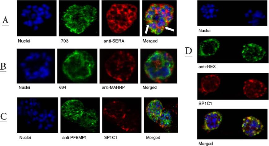
A. Immunofluorescence assay of rabbit antisera against PfMC-2TM peptides colocalized with anti-SERA 1 antibodies. Arrows indicate areas of colocalization between SERA 1 andPfMC-2TMB. Mouse antisera against MAHRP1 MAL13P1.413 was incubated with antisera against PfMC-2TM peptides.C. Immunofluorescence assay of rabbit antisera against PfEMP1 with Mabs SP1C1. PfEMP1 colocalized with PfMC-2TM, D. Immunofluorescence assay of rabbit antisera against REX1 PFI1735c colocalized with Mabs SP1C1. Optical sections were collected at a step size of 0.5 μm from the top to the bottom of the optical plane. Continuous overlap of the antibodies was observed.Tsarukyanova I, Drazba JA, Fujioka H, Yadav SP, Sam-Yellowe TY. Proteins of the Plasmodium falciparum two transmembrane Maurer's cleft protein family, PfMC-2TM, and the 130 kDa Maurer's cleft protein define different domains of the infected erythrocyte intramembranous network. Parasitol Res. 2009 104:875-91. Copyright Springer 2011.
See original on MMP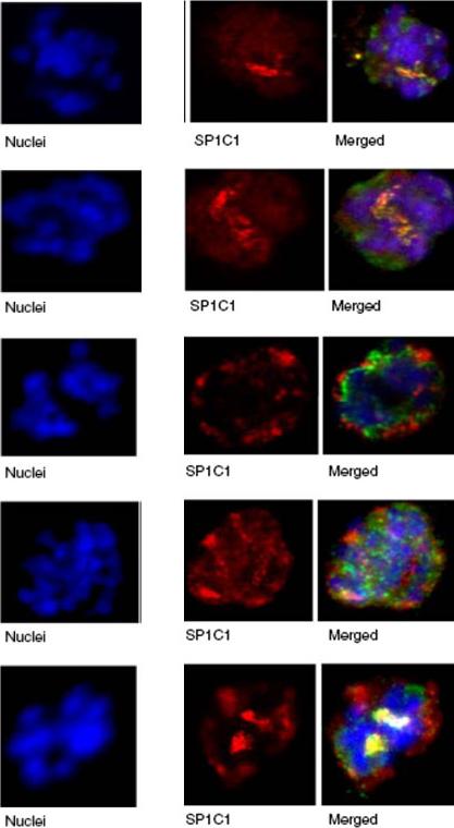
Immunofluorescence assay of rabbit antisera against PfMC-2TM peptides colocalized with Mab SP1C1. Immunofluorescence assays (IFA) in this figure and Figs. 4, 5, 6, 7, 8, and 9 were performed using the same stage of parasites. Trophozoite- and schizont-infected erythrocytes were incubated with mouse and rabbit primary antibodies, followed by secondary antibodies directed to both species, conjugated to different colored fluorochromes; Alexa 488 and Alexa 568 (Molecular Probes) for detection of primary antibodies. Antibody staining was considered colocalized if there was overlap (yellow/orange) in the staining pattern obtained with the two antibodies on or within the same structure or within domains of the intra-membranous network.Tsarukyanova I, Drazba JA, Fujioka H, Yadav SP, Sam-Yellowe TY. Proteins of the Plasmodium falciparum two transmembrane Maurer's cleft protein family, PfMC-2TM, and the 130 kDa Maurer's cleft protein define different domains of the infected erythrocyte intramembranous network. Parasitol Res. 2009 104:875-91. Copyright Springer 2011.
See original on MMP
Immunoelectron micrographs of young trophozoite-, late trophozoite-, young schizont-, and late schizont-infected erythrocytes reactive with rabbit antisera against PfMC-2TM peptides, 703, 705, and 713 (identical sequences in all PfMC-2TM-coding genes and Mab SP1A6 against the 130 kDa Maurer’s cleft protein. a–c Peptide-specific antisera are reactive with the PV/PVM and weakly around clefts. d–f Mab SP1A6 is reactive with structures within the parasite in young trophozoites, probably the endoplasmic reticulum (d), late trophozoites in erythrocyte cytoplasm under knobs (e), and in young schizonts around longitudinal clefts in the erythrocyte cytoplasm (f).Tsarukyanova I, Drazba JA, Fujioka H, Yadav SP, Sam-Yellowe TY. Proteins of the Plasmodium falciparum two transmembrane Maurer's cleft protein family, PfMC-2TM, and the 130 kDa Maurer's cleft protein define different domains of the infected erythrocyte intramembranous network. Parasitol Res. 2009 104:875-91. Copyright Springer 2011.
See original on MMP
A. Immunofluorescence assay of rabbit antisera against PfMC-2TM peptides colocalized with anti-SERA 1 antibodies. Arrows indicate areas of colocalization between SERA 1 andPfMC-2TMB. Mouse antisera against MAHRP1 MAL13P1.413 was incubated with antisera against PfMC-2TM peptides.C. Immunofluorescence assay of rabbit antisera against PfEMP1 with Mabs SP1C1. PfEMP1 colocalized with PfMC-2TM, D. Immunofluorescence assay of rabbit antisera against REX1 PFI1735c colocalized with Mabs SP1C1. Optical sections were collected at a step size of 0.5 μm from the top to the bottom of the optical plane. Continuous overlap of the antibodies was observed.Tsarukyanova I, Drazba JA, Fujioka H, Yadav SP, Sam-Yellowe TY. Proteins of the Plasmodium falciparum two transmembrane Maurer's cleft protein family, PfMC-2TM, and the 130 kDa Maurer's cleft protein define different domains of the infected erythrocyte intramembranous network. Parasitol Res. 2009 104:875-91. Copyright Springer 2011.
See original on MMP
Immunofluorescence assay of rabbit antisera against PfMC-2TM peptides colocalized with Mab SP1C1. Immunofluorescence assays (IFA) in this figure and Figs. 4, 5, 6, 7, 8, and 9 were performed using the same stage of parasites. Trophozoite- and schizont-infected erythrocytes were incubated with mouse and rabbit primary antibodies, followed by secondary antibodies directed to both species, conjugated to different colored fluorochromes; Alexa 488 and Alexa 568 (Molecular Probes) for detection of primary antibodies. Antibody staining was considered colocalized if there was overlap (yellow/orange) in the staining pattern obtained with the two antibodies on or within the same structure or within domains of the intra-membranous network.Tsarukyanova I, Drazba JA, Fujioka H, Yadav SP, Sam-Yellowe TY. Proteins of the Plasmodium falciparum two transmembrane Maurer's cleft protein family, PfMC-2TM, and the 130 kDa Maurer's cleft protein define different domains of the infected erythrocyte intramembranous network. Parasitol Res. 2009 104:875-91. Copyright Springer 2011.
See original on MMPMore information
| PlasmoDB | PF3D7_0101300 |
| GeneDB | PF3D7_0101300 |
| Malaria Metabolic Pathways | Localisation images Pathways mapped to |
| Previous ID(s) | MAL1P1.4, PFA0065w |
| Orthologs | |
| Google Scholar | Search for all mentions of this gene |