PBANKA_1432400 perforin-like protein 2 (PLP2)
Disruptability [+]
| Species | Disruptability | Reference | Submitter | |
|---|---|---|---|---|
| P. berghei ANKA |
Possible |
RMgm-839 | Imported from RMgmDB | |
| P. falciparum 3D7 |
Possible |
24602217 | Theo Sanderson, Wellcome Trust Sanger Institute | |
| P. falciparum 3D7 |
Possible |
USF piggyBac screen (Insert. mut.) | USF PiggyBac Screen | |
Mutant phenotypes [+]
| Species | Stage | Phenotype | Reference | Submitter |
|---|---|---|---|---|
| P. berghei ANKA | Asexual |
No difference |
RMgm-839 | Imported from RMgmDB |
| P. berghei ANKA | Gametocyte |
Difference from wild-type |
RMgm-839
Normal production of gametocytes. The production of male gametes is affected. The male gametocyte displays abnormal exflagellation; instead of forming 8 gametes, it produced only one, shared thicker flagellum. Evidence is presented that rupture of the erythrocyte membrane is blocked and thereby preventing egress of male gametes from the erythrocyte. |
Imported from RMgmDB |
| P. berghei ANKA | Ookinete |
Difference from wild-type |
RMgm-839
Strongly reduced ookinete production as a result of aberrant production of male gametes. The male gametocyte displays abnormal exflagellation; instead of forming 8 gametes, it produced only one, shared thicker flagellum. Evidence is presented that rupture of the erythrocyte membrane is blocked and thereby preventing egress of male gametes from the erythrocyte. Evidence is presented that female gametocytes/female gametes show normal egress from the erythrocyte. |
Imported from RMgmDB |
| P. berghei ANKA | Oocyst |
Difference from wild-type |
RMgm-839
Strongly reduced oocyst production as a result of strongly reduced ookinete production. |
Imported from RMgmDB |
| P. falciparum 3D7 | Asexual |
No difference |
24602217 | Theo Sanderson, Wellcome Trust Sanger Institute |
Imaging data (from Malaria Metabolic Pathways)
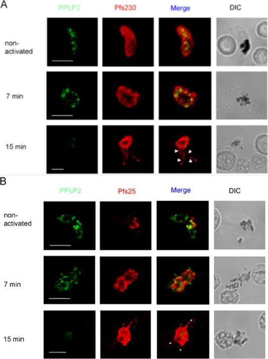
Location of PPLP2 in P. falciparum gametocytes during activation. A. Cultures of gametocytes before activation, and at 7 and 15 min post-activation, were labelled with antiserum B directed against PPLP2 (green) and counterstained with (A) anti-Pfs230 antisera (red) or (B) anti-Pfs25. The corresponding differential interference contrast (DIC) images are shown. Note that mature P. falciparum gametocytes have a banana-like shape but round up during activation. Bar, 5 μm (A) and 2 μm (B). Deligianni E, Morgan RN, Bertuccini L, Wirth CC, Silmon de Monerri NC, Spanos L, Blackman MJ, Louis C, Pradel G, Siden-Kiamos I. A perforin-like protein mediates disruption of the erythrocyte membrane during egress of Plasmodium berghei male gametocytes. Cell Microbiol. 2013 15(8):1438-55 PMID:
See original on MMP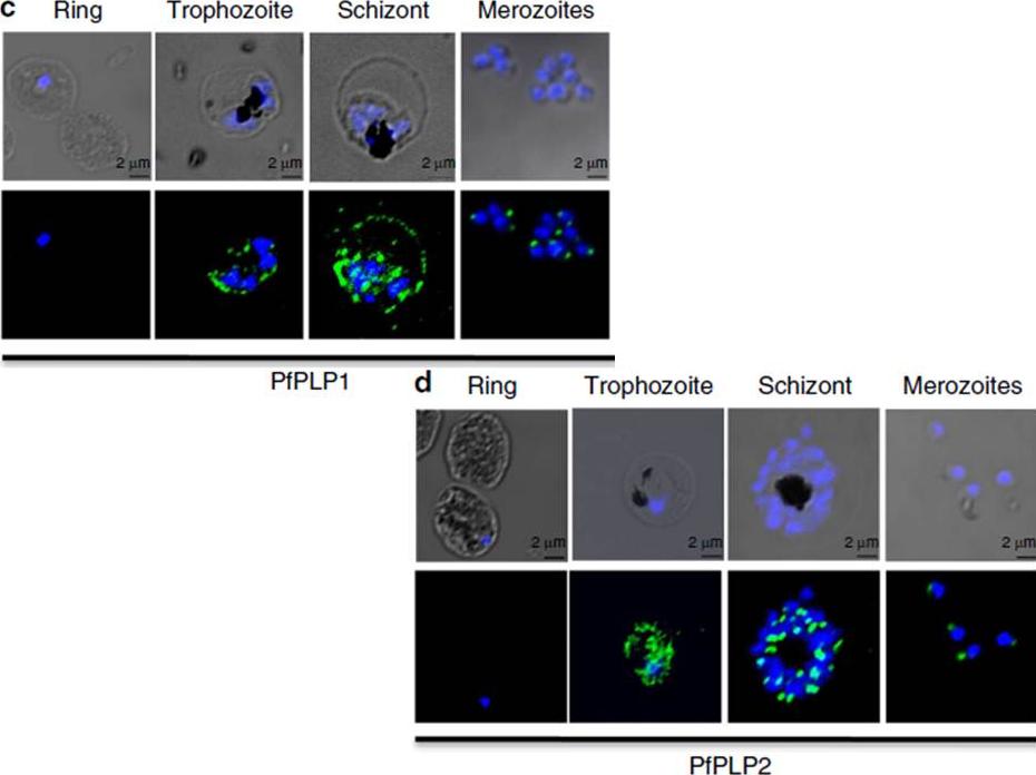
Stage-specific expression of PfPLP1 and PfPLP2 in the blood stage of P. falciparum. Expression of PfPLP1 and PfPLP2 starts in trophozoite stage, with maximal expression in schizonts. In mature schizonts, PfPLP1 and PfPLP2 show punctate staining at the apical end of merozoites. PfPLP1 also shows a ring-like staining, suggesting its localization to RBC membrane. Scale bar, 2 mm.Garg S, Agarwal S, Kumar S, Shams Yazdani S, Chitnis CE, Singh S. Calcium-dependent permeabilization of erythrocytes by a perforin-like protein during egress of malaria parasites. Nat Commun. 2013 Apr 16;4:1736.
See original on MMP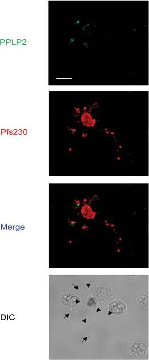
Immunofluorescence assays of P. falciparum gametocytes at 15 min post-activation showed PPLP2 labelling (green) in association with shed membranes (indicated by arrows). The exflagellating microgametocyte is highlighted by labelling of Pfs25 (red). Arrowheads indicate microgametes. The corresponding differential interference contrast (DIC) images are shown. Bar, 5 μm.Deligianni E, Morgan RN, Bertuccini L, Wirth CC, Silmon de Monerri NC, Spanos L, Blackman MJ, Louis C, Pradel G, Siden-Kiamos I. A perforin-like protein mediates disruption of the erythrocyte membrane during egress of Plasmodium berghei male gametocytes. Cell Microbiol. 2013 15(8):1438-55
See original on MMP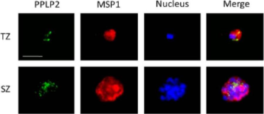
PPLP2 localization in asexual blood stage parasites. PPLP2-positive vesicular structures are present in the cytoplasm of trophozoites (TZ) and schizonts (SZ). PPLP2 was immunolabelled with mouse anti-PPLP2RP1 antisera (green), the asexual blood stages were visualized by rabbit anti-MSP1 antisera (red). Nuclei were highlighted by Hoechst stain (blue). PPLP2 first appeared in the trophozoites stage and here localized in vesicular structures. Multiple PPLP2-positive vesicular structures were detected also in mature schizonts. The blood stage parasites were highlighted by labelling of MSP1.Wirth CC, Glushakova S, Scheuermayer M, Repnik U, Garg S, Schaack D, Kachman MM, Weißbach T, Zimmerberg J, Dandekar T, Griffiths G, Chitnis CE, Singh S, Fischer R, Pradel G. Perforin-like protein PPLP2 permeabilizes the red blood cell membrane during egress of Plasmodium falciparum gametocytes. Cell Microbiol. 2014 Mar 7.
See original on MMPMore information
| PlasmoDB | PBANKA_1432400 |
| GeneDB | PBANKA_1432400 |
| Malaria Metabolic Pathways | Localisation images Pathways mapped to |
| Previous ID(s) | PB000619.01.0, PB001069.01.0, PBANKA_143240 |
| Orthologs | PCHAS_1434400 , PF3D7_1216700 , PKNH_1436300 , PVP01_1435500 , PVX_123515 , PY17X_1434700 |
| Google Scholar | Search for all mentions of this gene |