PBANKA_1421300 4-nitrophenylphosphatase, putative (PNPase)
Disruptability [+]
| Species | Disruptability | Reference | Submitter | |
|---|---|---|---|---|
| P. berghei ANKA |
Refractory |
https://www.biorxiv.org/content/early/2018/12/13/495473.1
Wrong integration of cassette, cytosolic localization |
Lakshmeesha K N et. al.,, JNCASR | |
| P. falciparum 3D7 |
Possible |
USF piggyBac screen (Insert. mut.) | USF PiggyBac Screen | |
Mutant phenotypes [+]
None reported yet. Please press the '+' button above to add one.Imaging data (from Malaria Metabolic Pathways)
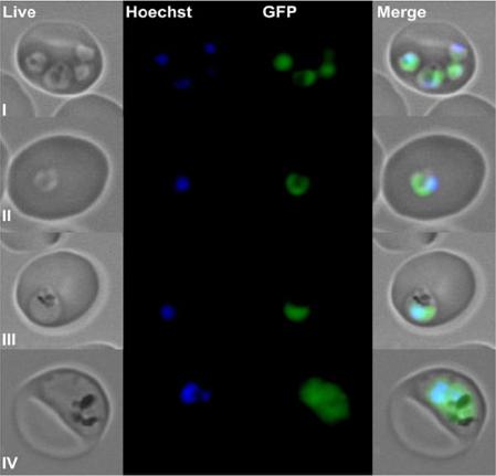
The open reading frame of the plasmodial PNPase was cloned in front of the GFP gene of the expression vector pARL1a− and subsequently transfected into P. falciparum. Developmental blood stages of the parasites were analysed by fluorescent light microscopy using an Axioskop 2 plus microscope. (I) Ring stage (multiple infection); (II) ring stage (single infection); (III) young trophozoite; (IV) old trophozoite. Live, P. falciparum live image; Hoechst, staining of the parasite’s nucleus; GFP, image taken using the GFP channel; Merge, merge of all images. The obtained fluorescent staining evidently proves the cytosolic appearance of the PfPNPase.Knöckel J, Bergmann B, Müller IB, Rathaur S, Walter RD, Wrenger C. Filling the gap of intracellular dephosphorylation in the Plasmodium falciparum vitamin B1 biosynthesis. Mol Biochem Parasitol. 2008 157:241-3. Copyright Elsevier 2009.
See original on MMPMore information
| PlasmoDB | PBANKA_1421300 |
| GeneDB | PBANKA_1421300 |
| Malaria Metabolic Pathways | Localisation images Pathways mapped to |
| Previous ID(s) | PB000763.02.0, PBANKA_142130 |
| Orthologs | PCHAS_1423100 , PF3D7_0715000 , PKNH_1424000 , PVP01_1424100 , PVX_122960 , PY17X_1423300 |
| Google Scholar | Search for all mentions of this gene |