PBANKA_0911700 subtilisin-like protease 2 (SUB2)
Disruptability [+]
| Species | Disruptability | Reference | Submitter | |
|---|---|---|---|---|
| P. berghei ANKA |
Refractory |
RMgm-160 | Imported from RMgmDB | |
| P. berghei ANKA |
Refractory |
PlasmoGEM (Barseq) | PlasmoGEM | |
| P. falciparum 3D7 |
Refractory |
15225303 | Theo Sanderson, Wellcome Trust Sanger Institute | |
| P. falciparum 3D7 |
Refractory |
USF piggyBac screen (Insert. mut.) | USF PiggyBac Screen | |
Mutant phenotypes [+]
| Species | Stage | Phenotype | Reference | Submitter |
|---|---|---|---|---|
| P. falciparum 3D7 | Asexual |
Invasion defect |
33287958 \"Here we show that genetic depletion of SUB2 abolishes shedding of a range of parasite proteins, identifying previously unrecognized SUB2 substrates. Interaction of SUB2-null merozoites with RBCs leads to either abortive invasion with rapid RBC lysis, or successful entry but developmental arrest. Selective failure to shed the most abundant SUB2 substrate, MSP1, reduces intracellular replication, whilst conditional ablation of the substrate AMA1 produces host RBC lysis. We conclude that SUB2 activity is critical for host RBC membrane sealing following parasite internalisation and for correct functioning of merozoite surface proteins.\" |
Theo Sanderson, Francis Crick Institute |
Imaging data (from Malaria Metabolic Pathways)
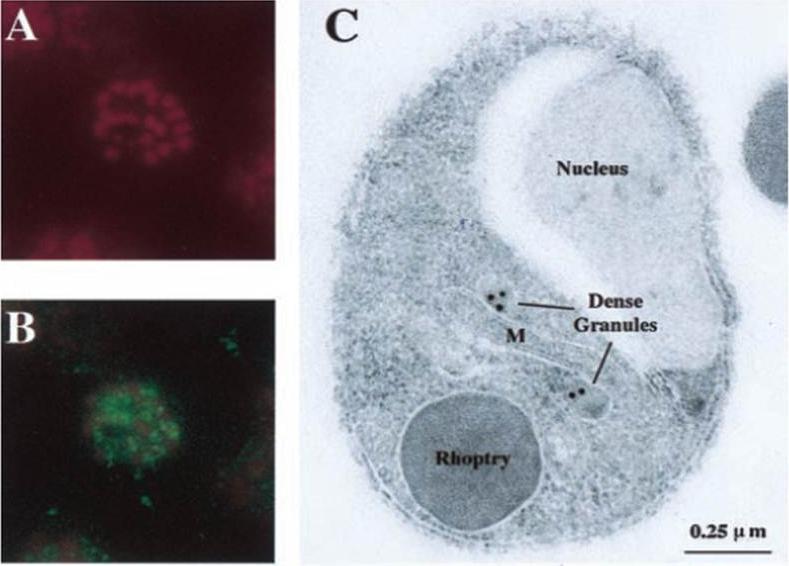
Localization of PfSUB2 protein by immunofluorescence on segmented schizonts and by IEM on fixed merozoites. (A and B) An air-dried fixed segmented schizont incubated with 25 mgyml of propidium iodide to label the parasite nuclei (A) and mouse anti-GSTPfSUB2 antibodies (B). Bound antibodies then were detected with FITC-coupled anti-mouse antibodies (magnification: 31,500). (C) The IEM analysis of thin sections of resin-embedded merozoites probed with mouse anti-GST-PfSUB2 antibodies and revealed with gold-labeled anti-mouse IgG antibody. M, mitochondrion.Barale JC, Blisnick T, Fujioka H, Alzari PM, Aikawa M, Braun-Breton C, Langsley G. Plasmodium falciparum subtilisin-like protease 2, a merozoite candidate for the merozoite surface protein 1-42 maturase. Proc Natl Acad Sci U S A. 1999 96:6445-50. Copyright 2009 National Academy of Sciences, U.S.A
See original on MMP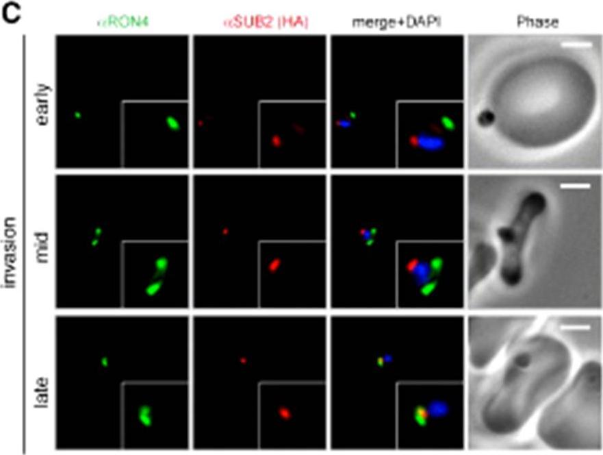
Wide field IFA time course of invasion using anti-HA(PfSUB2HA)/PfRON4. PfSUB2, is a micronemal protease, sheddase, responsible for MSP1 release. Scale bar = 2.0 μm. In parasites at early stages of invasion, the majority of PfSUB2 had reached the posterior pole of the merozoite (PfSUB2 early). Although not colocalized in the same parasite, the lack of contiguity between shed MSP1 and PfSUB2 sheddase, each in comparison with PfRON4 (PfSUB2 mid), suggests that MSP1 may be processed by PfSUB2 early in invasion but shed only during passage through the junction. In the latter stages of invasion, PfSUB2 remained at the posterior pole (SUB2 late).Riglar DT, Richard D, Wilson DW, Boyle MJ, Dekiwadia C, Turnbull L, Angrisano F, Marapana DS, Rogers KL, Whitchurch CB, Beeson JG, Cowman AF, Ralph SA, Baum J. Super-resolution dissection of coordinated events during malaria parasite invasion of the human erythrocyte. Cell Host Microbe. 2011 9:9-20.
See original on MMP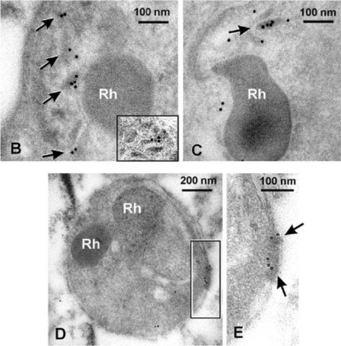
PfSUB2 Is a Microneme Protein. Electron micrographs showing immuno-gold labelling of micronemes within late-stage schizonts of PfSUB2HA clone 2D using: (B) anti-HA mAb 3F10, detecting epitope-tagged PfSUB2; and (C) a polyclonal antibody specific for PfAMA1. The inset in (B) shows another example of micronemal staining with mAb 3F10 from another schizont. Rh, rhoptry. (D and E) Posterior labelling of with anti-HA mAb 3F10 in a free merozoite of PfSUB2HA clone 2D. Arrows indicate immuno-gold labelling. Rh, rhoptry.Harris PK, Yeoh S, Dluzewski AR, O'Donnell RA, Withers-Martinez C, Hackett F, Bannister LH, Mitchell GH, Blackman MJ. Molecular identification of a malaria merozoite surface sheddase. PLoS Pathog. 2005 1(3):241-51.
See original on MMP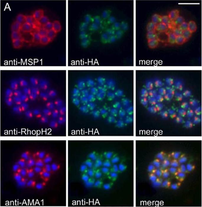
PfSUB2 Is a Microneme Protein. (A) IFA images of schizonts of PfSUB2HA clone 2D dual-labelled with mAbs X509 (anti-MSP1), 61.3 (anti-RhopH2), or 4G2 (anti-AMA1), plus in each case mAb 3F10 (anti-HA) The anti-HA signal co-localised only with the anti-AMA1 signal. Identical results were obtained with the uncloned transgenic PfSUB2HA line, and/or when anti-RAP2 mAb H5 was used as the rhoptry marker instead of mAb 61.3 (not shown). Parasite nuclei are stained throughout with DAPI (blue). Scale bar represents 2 lm.Harris PK, Yeoh S, Dluzewski AR, O'Donnell RA, Withers-Martinez C, Hackett F, Bannister LH, Mitchell GH, Blackman MJ. Molecular identification of a malaria merozoite surface sheddase. PLoS Pathog. 2005 1(3):241-51.
See original on MMP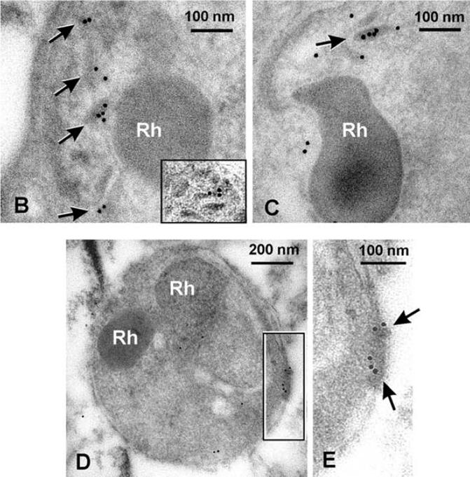
(B and C) Electron micrographs showing immuno-gold labelling of micronemes within late-stage schizonts of PfSUB2HA clone 2D using: (B) anti-HA mAb 3F10, detecting epitope-tagged PfSUB2; and (C) a polyclonal antibody specific for PfAMA1. The inset in (B) shows another example of micronemal staining with mAb 3F10 from another schizont. Rh, rhoptry. using the anti-HA mAb 3F10, which showed light but consistent labelling of typical elongated (D and E) Posterior labelling of with anti-HA mAb 3F10 in a free merozoite of PfSUB2HA clone 2D. Arrows indicate immuno-gold labelling. Rh, rhoptry. micronemes within late-stage schizonts (B), similar to that of AMA1 (C). No labelling with mAb 3F10 was observed in less-mature schizont stages lacking micronemes, or apically in free merozoites, and labelling was also absent from wild-type schizont controls (not shown).Harris PK, Yeoh S, Dluzewski AR, O'Donnell RA, Withers-Martinez C, Hackett F, Bannister LH, Mitchell GH, Blackman MJ. Molecular identification of a malaria merozoite surface sheddase. PLoS Pathog. 2005 Nov;1(3):241-51.
See original on MMP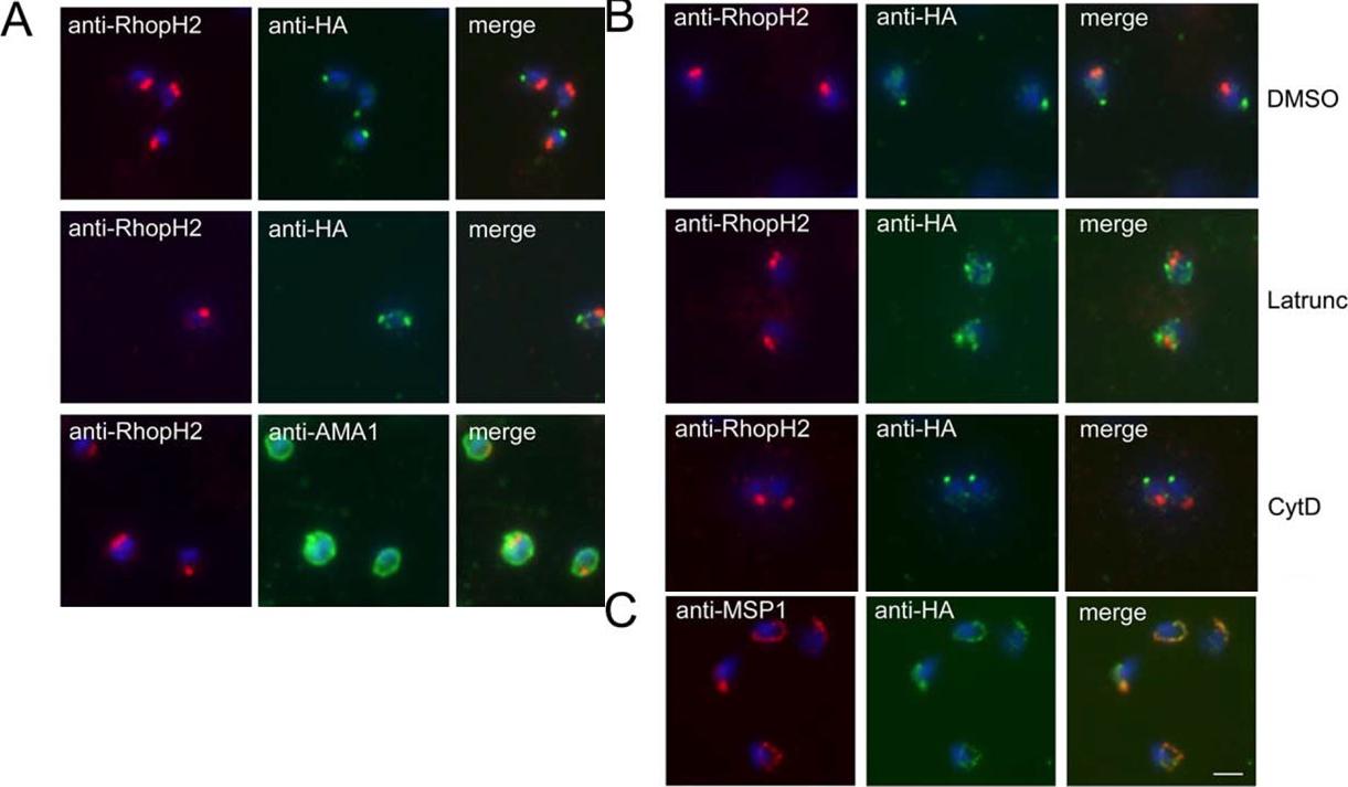
PfSUB2 Redistributes to the Free Merozoite Posterior in an Actin-Dependent Manner. (A) IFA images of free merozoites of PfSUB2HA clone 2D dual-labelled with mAb 61.3 (anti-RhopH2) and either mAb 3F10 (anti-HA) or 4G2 (anti-AMA1). The anti-HA signal formed either a single punctate signal at the extreme posterior of the merozoite (top), or twin foci lateral to its anterior–posterior axis (middle). Identical results were obtained using PfSUB2HA clone 10E, or using anti-RAP2 mAb H5 as the rhoptry marker (not shown). (B) Latrunculin inhibits rearward translocation of PfSUB2. Merozoites of PfSUB2HA clone 2D released into medium containing 1% (v/v) DMSO only (solvent control), or 5 mM latrunculin A (Latrunc), or 4 mM cytochalasin D (Cyt D), probed as above with mAb 61.3 (anti-RhopH2) and mAb 3F10 (anti-HA). Note that neither compound had any effect on the efficiency of schizont rupture and merozoite release, nor on AMA1 relocalisation, but both blocked erythrocyte invasion by more than 95%at the concentrations used (not shown). (C) PfSUB2 remains at the plasma membrane of the newly invaded parasite. Ring stages of PfSUB2HA clone 2D (<2 h post-invasion) probed with anti-MSP1 mAb 1E1 and anti-HA mAb 3F10. Parasite nuclei are stained throughout with DAPI (blue). Scale bar represents 2 mm.Harris PK, Yeoh S, Dluzewski AR, O'Donnell RA, Withers-Martinez C, Hackett F, Bannister LH, Mitchell GH, Blackman MJ. Molecular identification of a malaria merozoite surface sheddase. PLoS Pathog. 2005 Nov;1(3):241-51.
See original on MMP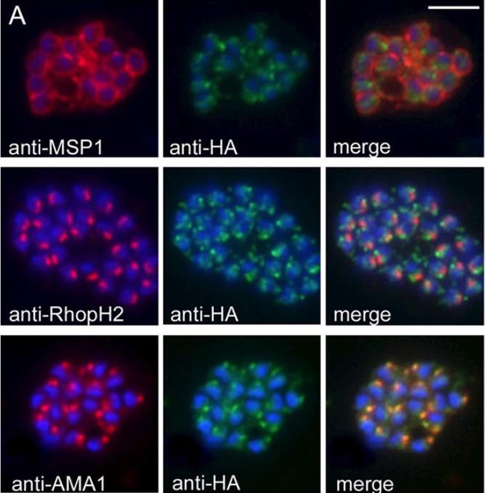
PfSUB2 Is a Microneme Protein. (A) IFA images of schizonts of PfSUB2HA clone 2D dual-labelled with mAbs X509 (anti-MSP1), 61.3 (anti-RhopH2), or 4G2 (anti-AMA1), plus in each case mAb 3F10 (anti-HA) The anti-HA signal co-localised only with the anti-AMA1 signal. Identical results were obtained with the unclonedtransgenic PfSUB2HA line, and/or when anti-RAP2 mAb H5 was used as the rhoptry marker instead of mAb 61.3 (not shown). Parasite nuclei are stained throughout with DAPI (blue). Scale bar represents 2 mm. the anti-HA signal was quite different from that of MSP1, it was adjacent to but distinct from that of the rhoptry specific signals, but it co-localised completely with that ofAMA1, indicating that in mature schizonts, PfSUB2 accumulates in micronemes.Harris PK, Yeoh S, Dluzewski AR, O'Donnell RA, Withers-Martinez C, Hackett F, Bannister LH, Mitchell GH, Blackman MJ. Molecular identification of a malaria merozoite surface sheddase. PLoS Pathog. 2005 Nov;1(3):241-51.
See original on MMPMore information
| PlasmoDB | PBANKA_0911700 |
| GeneDB | PBANKA_0911700 |
| Malaria Metabolic Pathways | Localisation images Pathways mapped to |
| Previous ID(s) | PB000680.03.0, PB300095.00.0, PB300857.00.0, PBANKA_091170 |
| Orthologs | PCHAS_0713000 , PF3D7_1136900 , PKNH_0935100 , PVP01_0937800 , PVX_092460 , PY17X_0913200 |
| Google Scholar | Search for all mentions of this gene |