PBANKA_0911500 apical exonemal protein, putative (AEP)
Disruptability [+]
| Species | Disruptability | Reference | Submitter | |
|---|---|---|---|---|
| P. berghei ANKA |
Possible |
PlasmoGEM (Barseq) | PlasmoGEM | |
| P. falciparum 3D7 |
Possible |
USF piggyBac screen (Insert. mut.) | USF PiggyBac Screen | |
Mutant phenotypes [+]
| Species | Stage | Phenotype | Reference | Submitter |
|---|---|---|---|---|
| P. berghei ANKA | Asexual |
Attenuated |
PlasmoGEM (Barseq) | PlasmoGEM |
Imaging data (from Malaria Metabolic Pathways)
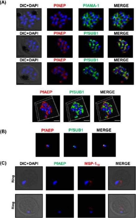
Localization of PfAEP in the exonemes of merozoites (A) Co-localization of PfAEP was studied with micronemal resident protein AMA-1 in bursting schizonts. PfAEP (rabbit serum; red) does not co-localize with AMA-1 (green). The staining for PfAEP co-localized with exonemal marker PfSUB1. Scale bar 2 mm. Three-dimensional reconstruction of the confocal z-stack images of schizonts is shown in panel below Scale bar 1.5mm (B) 2D-structured illumination microscopy (SIM) showed co-localization between the staining of PfAEP and PfSUB1 indicating its localization in the exonemes of P. falciparum merozoites. (C) Young rings were immunostained with anti PfAEP antibody and the antibody against MSP119 as a positive control. No staining was observed for PfAEP in young rings whereas MSP119 showed typical disk like staining around the nucleus.Hans N, Relan U, Dubey N, Gaur D, Chauhan VS. Identification and localization of a Novel Invasin of Plasmodium falciparum. Mol Biochem Parasitol. 2015 Sep 29. [Epub ahead of print]
See original on MMP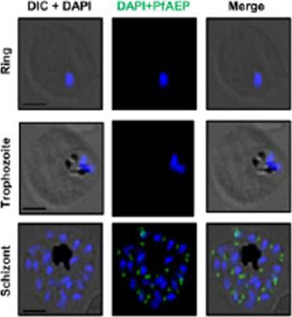
Expression of PfAEP was analyzed in different stages of the asexual blood-stage life cycle by confocal immuno-fluorescence microscopy using anti-PfAEP mouse serum (green). Parasite nuclei were counterstained with DAPI (blue). No detectable PfAEP staining was observed in ringand trophozoite stages. In schizonts, PfAEP was detected as a punctate staining characteristic of many apical organelle resident proteins. The scale bar indicates 2 mm. In schizonts, the staining of PfAEP was observed in a punctate manner towards the apex of individual merozoites similar to other invasion proteins.Hans N, Relan U, Dubey N, Gaur D, Chauhan VS. Identification and localization of a Novel Invasin of Plasmodium falciparum. Mol Biochem Parasitol. 2015 Sep 29. [Epub ahead of print] PMID:
See original on MMP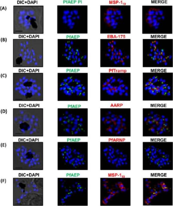
PfAEP localization in schizont stages analyzed by confocal microscopy. Subcellular localization of PfAEP was studied by co-staining with antibodies against micronemal protein EBA-175 (B), rhoptry bulb protein PTRAMP (C), and rhoptry neck,apical asparagine rich protein (AARP) (D), apical rhoptry neck protein (ARNP) (E), and merozoite surface protein (MSP) (F).As a control, pre-immune serum of PfAEP was checked and no staining was observed in schizonts (A). Mature schizonts were costained with anti-PfAEP (green) and anti-EBA-175, TRAMP, AARP, ARNP, MSP-119 antibodies (red). The nuclei of schizonts were stained with DAPI (blue) and visualized by Nikon N-SIM confocal microscope. All apical marker proteins and PfAEP showed punctate staining in schizonts distinct from DAPI. Co-staining of PfAEP with surface marker MSP-119 showed PfARNP localized at the apical tip with surface of merozoites stained by MSP-119. PfAEP staining did not merge with either markers of microneme or rhoptry indicating that PfAEP is neither a resident of micronemes or rhoptry ofmerozoites. Scale bar 2 mm.Hans N, Relan U, Dubey N, Gaur D, Chauhan VS. Identification and localization of a Novel Invasin of Plasmodium falciparum. Mol Biochem Parasitol. 2015 Sep 29. [Epub ahead of print]
See original on MMPMore information
| PlasmoDB | PBANKA_0911500 |
| GeneDB | PBANKA_0911500 |
| Malaria Metabolic Pathways | Localisation images Pathways mapped to |
| Previous ID(s) | PB000815.02.0, PB300806.00.0, PBANKA_091150 |
| Orthologs | PCHAS_0712800 , PF3D7_1137200 , PKNH_0935400 , PVP01_0938100 , PVX_092470 , PY17X_0912900 |
| Google Scholar | Search for all mentions of this gene |