PBANKA_0903600 dynamin-like protein, putative (DYN1)
Disruptability [+]
| Species | Disruptability | Reference | Submitter | |
|---|---|---|---|---|
| P. berghei ANKA |
Refractory |
RMgm-238 | Imported from RMgmDB | |
| P. falciparum 3D7 |
Refractory |
USF piggyBac screen (Insert. mut.) | USF PiggyBac Screen | |
Mutant phenotypes [+]
None reported yet. Please press the '+' button above to add one.Imaging data (from Malaria Metabolic Pathways)
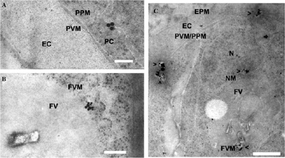
Location of Pfdyn1 in P. falciparum-infected erythrocytes with transmission electron. Percoll-synchonized late stage cells were fixed, and then incubated with purified anti-Pfdyn1-C antibody (1:100). Then incubated for in 15nm gold labeled goat anti-rabbit IgG diluted 1:20 in PBT and stained with uranyl acetate and lead citrate. FV, food vacuole; FVM, food vacuole membrane; EC, erythrocyte cytoplasm; EPM, erythrocyte plasma membrane; PC, parasite cytoplasm; PVM, parasitophorous vacuole membrane; PPM, parasite plasma membrane; N, nuclear; and NM, nuclear membrane. Bar, 100nm in (A), 200nm in (B), and 500nm in (C). Vesicles containing Pfdyn1-associated vesicles are indicated by arrowheads (>). The immunogold particles were observed in the cytoplasm of parasite and associated with membrane-like organelles such as food vacuoles, nucleus membrane, and some other unconfirmed membrane structures. Those particles were also seen in the cytoplasm of erythrocyte and on the parasite plasma membrane (PPM), parasitophorous vacuole membrane (PVM), and even erythrocyte plasma membrane (EPM).Li H, Han Z, Lu Y, Lin Y, Zhang L, Wu Y, Wang H. Isolation and functional characterization of a dynamin-like gene from Plasmodium falciparum. Biochem Biophys Res Commun. 2004 320:664-71. Copyright Elsevier 2011
See original on MMP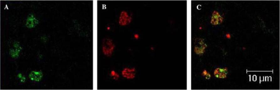
Location of Pfdyn1 in P. falciparum-infected erythrocytes by immunofluorescence microscopy. Percoll-synchronized late stage cells were smeared onto coverslips and fixed in acetone for 10 min at room temperature. The smears were probed with purified rabbit anti-Pfdyn1-C antibody (1:300) and rat anti-PfGRP antiserum (1:500). Binding of rabbit antibodies was visualized using goat anti-rabbit IgG conjugated with rhodamine. Binding of rat antibodies was visualized using goat anti-rat IgG conjugated with FITC. Coverslips were viewed under Leica TCS NT laser confocal scanning microscope. (A) FITC fluorescent; (B) rhodamine fluorescent; and (C) merged. The location of Pfdyn1 partially overlapped with that of PfGRP PFI0875w, an ER-resident protein. There iss a partial overlap of intracellular localization between Pfdyn1 and PfGRP.Li H, Han Z, Lu Y, Lin Y, Zhang L, Wu Y, Wang H. Isolation and functional characterization of a dynamin-like gene from Plasmodium falciparum. Biochem Biophys Res Commun. 2004 320:664-71. Copyright Elsevier 2011.
See original on MMP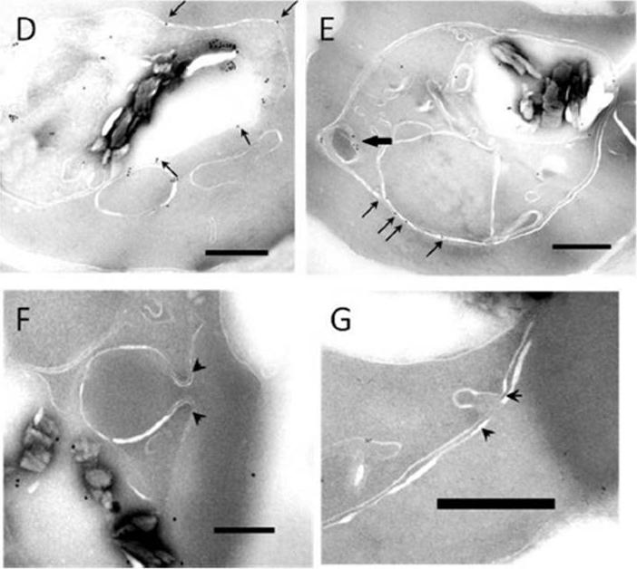
Immunoelectron micrographs showing intraparasitic localization of FKBP-HA-tagged P. falciparum Dyn1. The arrows indicate gold particle labeling on membranous structures. (E) Black arrow indicating HCs labeling. (F and G) Representative images showing that P. falciparum Dyn1 is absent from cytostomal collars (arrowheads). Scale bar, 250 nm.Milani KJ, Schneider TG, Taraschi TF. Defining the Morphology and Mechanism of the Hemoglobin Transport Pathway in Plasmodium falciparum-Infected Erythrocytes. Eukaryot Cell. 2015 14(4):415-26. PMID:
See original on MMPMore information
| PlasmoDB | PBANKA_0903600 |
| GeneDB | PBANKA_0903600 |
| Malaria Metabolic Pathways | Localisation images Pathways mapped to |
| Previous ID(s) | PB000159.03.0, PBANKA_090360 |
| Orthologs | PCHAS_0704800 , PF3D7_1145400 , PKNH_0943300 , PVP01_0946100 , PVX_092875 , PY17X_0905000 |
| Google Scholar | Search for all mentions of this gene |