PBANKA_0832000 selenoprotein, putative (Sel2)
Disruptability [+]
| Species | Disruptability | Reference | Submitter | |
|---|---|---|---|---|
| P. berghei ANKA |
Possible |
PlasmoGEM (Barseq) | PlasmoGEM | |
| P. falciparum 3D7 |
Possible |
USF piggyBac screen (Insert. mut.) | USF PiggyBac Screen | |
Mutant phenotypes [+]
| Species | Stage | Phenotype | Reference | Submitter |
|---|---|---|---|---|
| P. berghei ANKA | Asexual |
No difference |
PlasmoGEM (Barseq) | PlasmoGEM |
Imaging data (from Malaria Metabolic Pathways)
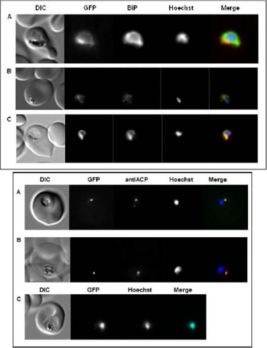
Upper panel: Subcellular localization of PfSel1, PfSel2, and PfSel4 in P. falciparum using GFP-fusion proteins. Shown are erythrocytes infected with trophozoite stage parasites. A. In the different Sel genes the UGA codon was replaced with different codons. PfSel1 (Sec->Cys), B. PfSel2 (Sec->Cys), and C. PfSel4 (Sec->Cys). Anti-BiP and Hoechst 33258 were used for co-localization with ER and nucleus, respectively. All gene products localized to the ER.Lower panel: Subcellular localization of PfSel3 in P. falciparum using GFP-fusion proteins and different PfSel3 mutants. Shown are erythrocytes infected with trophozoite stage parasites. A. PfSel3 (Sec->Cys), B. PfSel3 (Sec->Trp), C. PfSel3-UGA. Anti-ACP and Hoechst 33258 were used for co-localization with apicoplast and nucleus, respectively. PfSel3/GFP-fusion protein, which contains its original UGA codon, localized to the nucleus. All other proteins where the UGA codon has been replaced with different codons, localized to the apicoplast.Roeseler A, Prieto JH, Iozef R, Hecker B, Schirmer H, Kuelzer S, Przyborski J, Rahlfs S, Becker K. Insight into the selenoproteome of the malaria parasite Plasmodium falciparum. Antioxid Redox Signal. 2012 Jan 9. [Epub ahead of print]
See original on MMP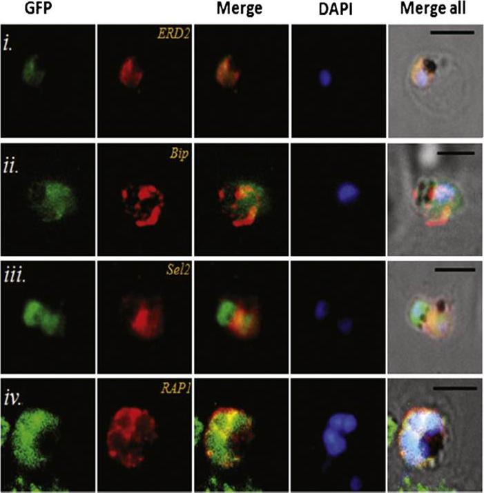
Transgenic parasites expressing Pfμ1–GFP were treated with Brefeldin A (BFA) and immunostained with antibodies specific to cis-Golgi apparatus marker ERD2 (i), endoplasmic reticulum marker, Bip (ii), cytoplasm localized, Sel2 (iii) and RAP1 (iv). The Pfμ1–GFP fusion protein colocalized with Sel2 as well as ERD2 in the parasite cytoplasm upon BFA treatment (i & iii). Parasite nuclei were stained with DAPI; scale bars denote 5 μm. Co-localization with antibodies to ERD2 (a cis-Golgi marker) and BiP, as well as the resident rhoptry protein RAP1 and the cytosolic protein Sel2 showed that Pfμ1 did not show a similar pattern as Bip or Sel2Pfμ1 showed a substantially similar staining pattern as ERD2 which is a Golgi marker (i–iii). Pfμ1 also did co-localize with RAP1 in BFA treated parasites (iv). These observations are consistent with Pfμ1 being Golgi associated at this life stage, consistent with its similar dynamics of redistribution upon brefeldin treatment.Kaderi Kibria KM, Rawat K, Klinger CM, Datta G, Panchal M, Singh S, Iyer GR, Kaur I, Sharma V, Dacks JB, Mohmmed A, Malhotra P. A role for adaptor protein complex 1 in protein targeting to rhoptry organelles in Plasmodium falciparum. Biochim Biophys Acta. 2015 1853(3):699-710.
See original on MMPMore information
| PlasmoDB | PBANKA_0832000 |
| GeneDB | PBANKA_0832000 |
| Malaria Metabolic Pathways | Localisation images Pathways mapped to |
| Previous ID(s) | PBANKA_083200, Sel2 |
| Orthologs | PCHAS_0832300 , PF3D7_0931200 , PKNH_0729900 , PVP01_0729900 , PVX_087143 , PY17X_0835400 |
| Google Scholar | Search for all mentions of this gene |