PBANKA_0813000 inhibitor of cysteine proteases (ICP)
Disruptability [+]
| Species | Disruptability | Reference | Submitter | |
|---|---|---|---|---|
| P. berghei ANKA |
Possible |
RMgm-1114 | Imported from RMgmDB | |
| P. berghei ANKA |
Possible |
RMgm-970 | Imported from RMgmDB | |
| P. berghei ANKA |
Refractory |
PlasmoGEM (Barseq) | PlasmoGEM | |
| P. falciparum 3D7 |
Possible |
USF piggyBac screen (Insert. mut.) | USF PiggyBac Screen | |
Mutant phenotypes [+]
| Species | Stage | Phenotype | Reference | Submitter |
|---|---|---|---|---|
| P. berghei ANKA | Asexual |
Difference from wild-type |
RMgm-1114
Asexual blood infections in mice induced infections with a delayed increase of the parasitemia |
Imported from RMgmDB |
| P. berghei ANKA | Asexual |
No difference |
RMgm-970 | Imported from RMgmDB |
| P. berghei ANKA | Gametocyte |
No difference |
RMgm-1114 | Imported from RMgmDB |
| P. berghei ANKA | Ookinete |
No difference |
RMgm-1114 | Imported from RMgmDB |
| P. berghei ANKA | Oocyst |
No difference |
RMgm-1114 | Imported from RMgmDB |
| P. berghei ANKA | Oocyst |
No difference |
RMgm-970 | Imported from RMgmDB |
| P. berghei ANKA | Sporozoite |
Difference from wild-type |
RMgm-1114
Normal numbers of oocyst-derived (hemocoel) sporozoites are formed. Sporozoites have greatly impaired gliding motility and capacity to invade salivary glands. |
Imported from RMgmDB |
| P. berghei ANKA | Sporozoite |
Difference from wild-type |
RMgm-970
Only 'hemocoel-associated' sporozoites are formed. Sporozoites do not invade salivary glands. Hemocoel sporozoites show reduced gliding. Sporozoites are not infectious to hepatocytes in culture. |
Imported from RMgmDB |
| P. berghei ANKA | Liver |
Difference from wild-type |
RMgm-1114
Normal numbers of oocyst-derived (hemocoel) sporozoites are formed. Sporozoites have greatly impaired gliding motility and capacity to invade salivary glands. Sporozoites are not able to invade hepatocytes in vitro. Sporozoites were not infectious to mice. |
Imported from RMgmDB |
| P. berghei ANKA | Liver |
Difference from wild-type |
RMgm-970
Sporozoites are not infectious to hepatocytes in culture and not infectious to mice (after intravenous inoculation of hemocoel sporozoites) |
Imported from RMgmDB |
Imaging data (from Malaria Metabolic Pathways)
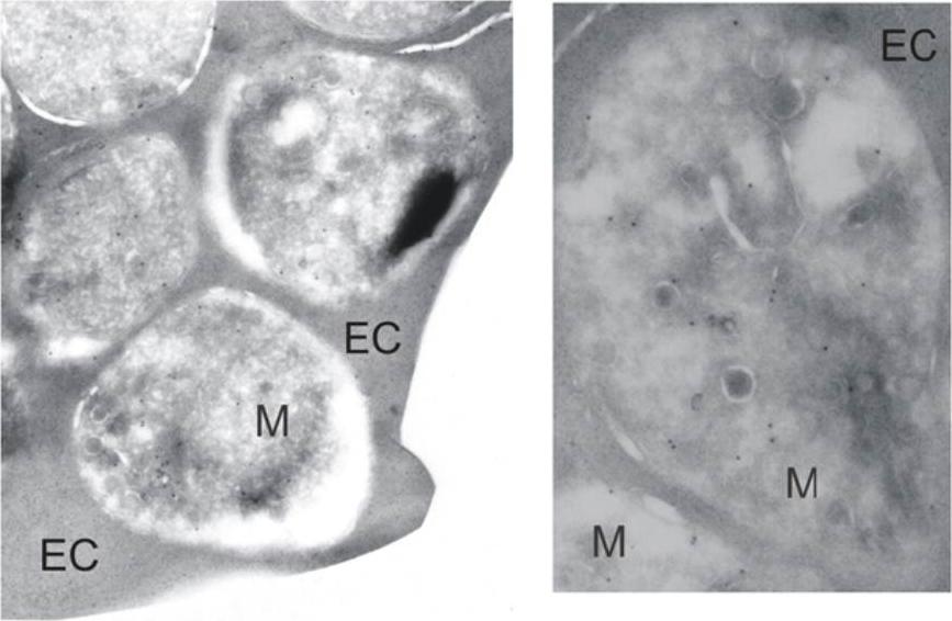
Immunoelectron microscopy. Late-schizont stage parasites were incubated with anti-falstatin antibodies and gold-conjugated second antibody and then evaluated by electron microscopy. Labels show individual merozoites (M) and erythrocyte cytosol (EC). Falstatin localizes to the periphery of rings and early schizonts, is diffusely expressed in late schizonts and merozoites, and is released upon the rupture of mature schizonts.Pandey KC, Singh N, Arastu-Kapur S, Bogyo M, Rosenthal PJ. Falstatin, a cysteine protease inhibitor of Plasmodium falciparum, facilitates erythrocyte invasion. PLoS Pathog. 2006 2(11):e117.
See original on MMP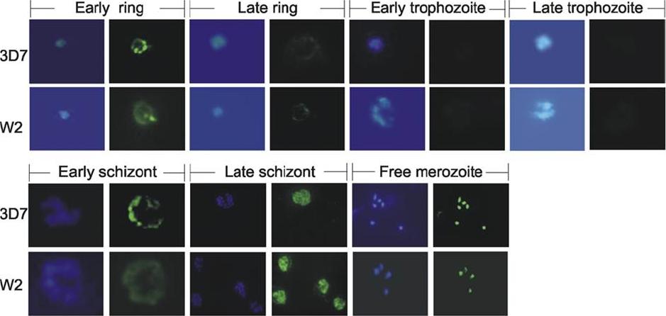
Immunolocalization of Falstatin. Immunofluorescence microscopy. Erythrocytes infected with synchronized 3D7 or W2 parasites were collected every 8 h, stained with DAPI and anti-falstatin antibodies and FITC-second antibody, and then evaluated by immunofluorescence microscopy. Falstatin is present in schizonts, merozoites, and rings, but not in trophozoites, the stage at which the cysteine protease activity of P. falciparum is maximal. Pandey KC, Singh N, Arastu-Kapur S, Bogyo M, Rosenthal PJ. Falstatin, a cysteine protease inhibitor of Plasmodium falciparum, facilitates erythrocyte invasion. PLoS Pathog. 2006 2(11):e117.
See original on MMPMore information
| PlasmoDB | PBANKA_0813000 |
| GeneDB | PBANKA_0813000 |
| Malaria Metabolic Pathways | Localisation images Pathways mapped to |
| Previous ID(s) | PB000502.02.0, PBANKA_081300 |
| Orthologs | PCHAS_0813300 , PF3D7_0911900 , PKNH_0709900 , PVP01_0710400 , PVX_099035 |
| Google Scholar | Search for all mentions of this gene |