PBANKA_0713900 adenylate kinase 2, putative (AK2)
Disruptability [+]
| Species | Disruptability | Reference | Submitter | |
|---|---|---|---|---|
| P. berghei ANKA |
Possible |
PlasmoGEM (Barseq) | PlasmoGEM | |
| P. falciparum 3D7 |
Refractory |
USF piggyBac screen (Insert. mut.) | USF PiggyBac Screen | |
Mutant phenotypes [+]
| Species | Stage | Phenotype | Reference | Submitter |
|---|---|---|---|---|
| P. berghei ANKA | Asexual |
No difference |
PlasmoGEM (Barseq) | PlasmoGEM |
Imaging data (from Malaria Metabolic Pathways)
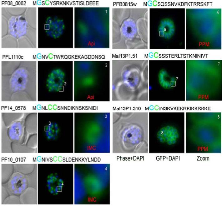
Localization of the predicted N-terminal (20aa) palmitoylated and myristoylated proteins in P. falciparum. The prediected peptides (indicated above the micrographs) were localized in late stage parasite using GFP fusion proteins. According to their predominat subcellular distribution they were grouped into apical (Api), inner membrane complex (IMC) and paraiste plasma membrane (PPM). Nuclei stained with DAPI. Enlargement of selected areas are marked with white square and referred as Zoom. Scale bar, 1 μm.Rahlfs S, Koncarevic S, Iozef R, Mailu BM, Savvides SN, Schirmer RH, Becker K. Myristoylated adenylate kinase-2 of Plasmodium falciparum forms a heterodimer with myristoyltransferase. Mol Biochem Parasitol. 2009 163(2):77-84 PMID: . Copyright Elsevier
See original on MMP
Localization of PfAK2. (A) Co-localization of PfAK2-GFP with serine-rich protein (SERA5). (B) A comparison the localization of PfAK2-GFP with Bodipy-TR-ceramide shows that the loops contain membranous material. (C) Co-localization of PfAK2-GFP with Plasmodium falciparum exported protein (Exp1) by using an anti-Exp1 antibody. Scale bar, 3 mm. PfAK2 localized to a ring-like structure around the 264 parasite, and ‘‘loops’’ apparently connected to the PVM. PfAK2-GFP co localizes with the PV resident protein SERA5 which seems to be excluded from the ‘‘loops’’ suggesting that PfAK2-GFP in actual fact associates with the membrane of the PV, rather than being found in a soluble state in the PV lumen.Ma J, Rahlfs S, Jortzik E, Heiner Schirmer R, Przyborski J, Becker K. Subcellular localization of adenylate kinases in Plasmodium falciparum. FEBS Lett. 2012 Jul 17. [Epub ahead of print]
See original on MMP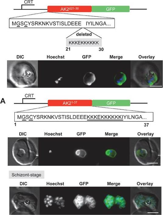
Upper panel: Deletion of the stretch of basic residues at the N-terminus of PfAK2 alters the subcellular location of the protein. (A) The AK2Δ21-30/GFP was expressed using the CRT promoter (construct indicated above the images). Live cell imaging of the PfAK2Δ21-30/GFP parasite line in a late trophozoite stage parasite.Lower panel: The PfAK2 N-terminus targets GFP to the outside of the parasite plasma membrane. (A) The AK21-37/GFP was expressed using the CRTpromoter (construct indicated above the images). Live cell imaging of the PfAK21-37/GFP in trophozoite and schizont stages, showed a location of the GFP signal similar to that of PfAK2/GFP. In the schizont stage of these parasites a clear signal for the fusion protein was visible around each of the individualdaughter merozoites.Thavayogarajah T, Gangopadhyay P, Rahlfs S, Becker K, Lingelbach K, Przyborski JM, Holder AA. Alternative Protein Secretion in the Malaria Parasite Plasmodium falciparum. PLoS One. 2015 Apr 24;10(4):e0125191.
See original on MMP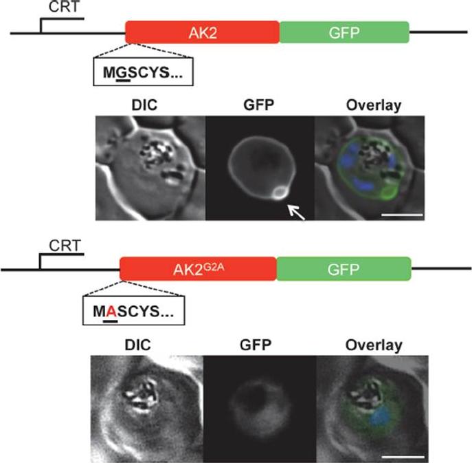
Subcellular location of PfAK2/GFP and the non-myristoylated G2A variant, as determined by live fluorescence microscopy (A) The AK2/GFP and the AK2G2A/GFP fusion proteins were expressed using the CRT promoter from an episomal plasmid (constructs indicated above the images). AK2/GFP is located at the periphery of the intracellular parasite as judged by epifluorescence microscopy, and is associated with one or two protuberances towards the host cell cytoplasm present on each parasite (indicated by white arrow). In contrast, the AK2G2A/GFP chimera is located within the parasite cytosol. The infected cell was visualised by differential interference contrast (DIC), intrinsic fluorescence of the GFP identified the location of the AK2/GFP fusion protein, and parasite nuclei were detected by Hoechst staining. Overlay: green (GFP), blue (DNA). Scale bar-3 μm.Thavayogarajah T, Gangopadhyay P, Rahlfs S, Becker K, Lingelbach K, Przyborski JM, Holder AA. Alternative Protein Secretion in the Malaria Parasite Plasmodium falciparum. PLoS One. 2015 Apr 24;10(4):e0125191.
See original on MMP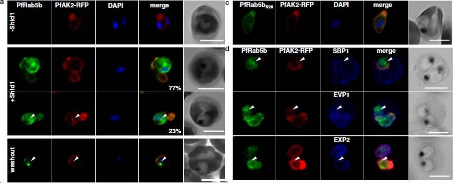
PfRab5b expression disturbed the transport of PfAK2 to the TVN. a In the absence of Shld1, PfAK2-RFP (red) localized to the TVN, while PfRab5b-YFP-DD signal was not detect (−Shld1, upper panel). After Shld1 stabilization for 48 h, PfRab5b-YFP-DD (green) and PfAK2-RFP co-localized in the TVN-like structures (+Shld1, middle-upper panel). In 77 % of PfRab5b-YFP-DD-positive parasites, PfAK2-RFP localized to the TVN, while in 23 % of PfRab5b-YFP-DD positive parasites, PfAK2-RFP signals also accumulated in the punctate compartment within the parasite (+Shld1, middle-lowerpanel, arrowhead). Co-localization of PfRab5b-YFP-DD and PfAK2-RFP at the punctate compartment remained even after removal of Shld1 (washout, lower panel, arrowhead). c No punctate localization of PfAK2-RFP was apparent with co-expression of PfRab5bN20-YFP-DD. d Effect of PfRab5b over-expression on the exported proteins PfSBP1, PfEVP1, and PfEXP2. In contrast to PfAK2-RFP, these proteins did not accumulate in the PfRab5b- and PfAK2-double positive punctate compartments (arrowhead).Ebine K, Hirai M, Sakaguchi M, Yahata K, Kaneko O, Saito-Nakano Y. Plasmodium Rab5b is secreted to the cytoplasmic face of the tubovesicular network in infected red blood cells together with N-acylated adenylate kinase 2. Malar J. 2016 15:323.
See original on MMPMore information
| PlasmoDB | PBANKA_0713900 |
| GeneDB | PBANKA_0713900 |
| Malaria Metabolic Pathways | Localisation images Pathways mapped to |
| Previous ID(s) | PB000875.00.0, PB300697.00.0, PBANKA_071390 |
| Orthologs | PCHAS_0723000 , PF3D7_0816900 , PKNH_0503500 , PVP01_0518400 , PVX_089565 , PY17X_0714100 |
| Google Scholar | Search for all mentions of this gene |