PBANKA_0702800 protein disulfide isomerase
Disruptability [+]
| Species | Disruptability | Reference | Submitter | |
|---|---|---|---|---|
| P. berghei ANKA |
Refractory |
PlasmoGEM (Barseq) | PlasmoGEM | |
| P. falciparum 3D7 |
Refractory |
USF piggyBac screen (Insert. mut.) | USF PiggyBac Screen | |
Mutant phenotypes [+]
None reported yet. Please press the '+' button above to add one.Imaging data (from Malaria Metabolic Pathways)
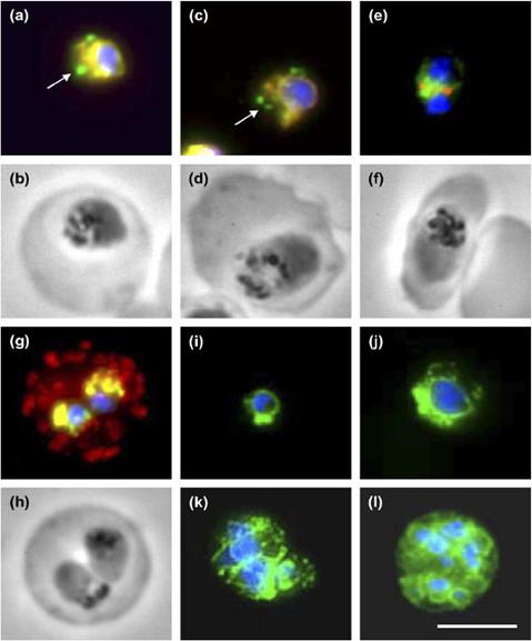
PfPDI localization in different parasite erythrocytic stages and co-localization with markers of subcellular compartments. (a,b) Immunostaining with antisera directed against rPfPDI (green) and PfBIP-PFI0875w (red) and the corresponding phase contrast image of a trophozoite. (c,d) Immunostaining with antisera directed against rPfPDI (green) and PfERC-PF11_0098 (red) and the corresponding phase contrast image of a trophozoite. (e,f) Immunostaining with antisera directed against rPfPDI (green) and PfERD2-PF13_0280 (red) and the corresponding phase contrast image of a young schizont. (g,h) Immunostaining with antisera directed against rPfPDI (green) and Pf332-PF11_0507 (red) and the corresponding phase contrast image of an erythrocyte infected by two young trophozoites. Regions of total overlapping of both proteins appear a yellow-orange color, DNA staining by DAPI appears in blue color. Arrows indicate the absence of co-localization. (i-l) Overlaid images of parasites immunostained with antiserum directed against rPfPDI (green) and incubated with the nuclear dye DAPI (blue). Parasite stages were: ring (i), old trophozoites (j), schizont (k) and mature schizont with individualized merozoites (l). Bar: 5 mm. PfPDI with endoplasmic reticulum-resident proteins, PfBIP and PfERC, but not with the Golgi marker PfERD2 or the Maurer’s cleft marker Pf332-PF11_0507.Mouray E, Moutiez M, Girault S, Sergheraert C, Florent I, Grellier P. Biochemical properties and cellular localization of Plasmodium falciparum protein disulfide isomerase. Biochimie. 2007 89:337-46. Copyright Elsevier 2009.
See original on MMP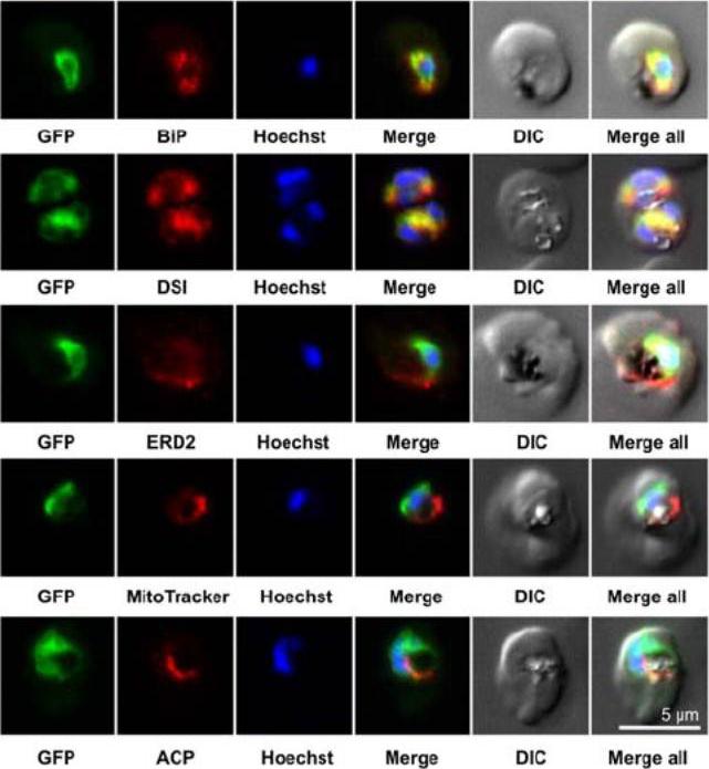
Colocalization studies. PfSCD-GFP was colocalized with different compartment markers: ER (PfBip, PfDSI), cis-Golgi (PfERD2), mitochondria (MitoTracker Red), and apicoplast (PfACP PFB0385w). PfSCD-GFP colocalized mostly with PfBiP and PfDSI (disulfide isomerase), two soluble ER markers, therefore confirming the ER localization of PfSCD-GFP. In contrast, the GFP-tagged PfSCD protein did not colocalize with ERD2, a known marker of the cis-Golgi.Gratraud P, Huws E, Falkard B, Adjalley S, Fidock DA, Berry L, Jacobs WR Jr, Baird MS, Vial H, Kremer L. Oleic acid biosynthesis in Plasmodium falciparum: characterization of the stearoyl-CoA desaturase and investigation as a potential therapeutic target. PLoS One. 2009 4(9):e6889.
See original on MMPMore information
| PlasmoDB | PBANKA_0702800 |
| GeneDB | PBANKA_0702800 |
| Malaria Metabolic Pathways | Localisation images Pathways mapped to |
| Previous ID(s) | PB001298.00.0, PBANKA_070280 |
| Orthologs | PCHAS_0935200 , PF3D7_0827900 , PKNH_1322000 , PVP01_0506500 , PVX_088960 , PY17X_0703100 |
| Google Scholar | Search for all mentions of this gene |