PBANKA_0602000 origin recognition complex subunit 1, putative (ORC1)
Disruptability [+]
| Species | Disruptability | Reference | Submitter | |
|---|---|---|---|---|
| P. berghei ANKA |
Refractory |
PlasmoGEM (Barseq) | PlasmoGEM | |
| P. falciparum 3D7 |
Possible |
USF piggyBac screen (Insert. mut.) | USF PiggyBac Screen | |
Mutant phenotypes [+]
None reported yet. Please press the '+' button above to add one.Imaging data (from Malaria Metabolic Pathways)
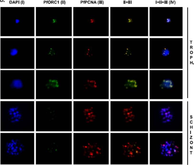
Colocalization of PfPCNA1 and PfORC1 during the replicating-trophozoite stage. A glass slide containing a parasite smear from the replicating-trophozoite stage or schizont stage was treated for immunofluoresence studies as described in Materials and Methods using both anti-PfORC1 and anti-PfPCNA1 antibodies. Distinct colocalization of PfORC1 and PfPCNA1 foci can be detected during the replicating-trophozoite stage (Troph.; rows 1 to 3). Although the expression of PfPCNA can be detected at the multinucleated late schizont stage (rows 4 and 5), the expression of PfORC1 is barely visible as the protein is degraded at the late stage, as described earlier. DAPI (4’,6-diamidino-2-phenylindole) (I) shows the nuclei. Column IV represents merged panels I, II, and III. Gupta A, Mehra P, Deshmukh A, Dar A, Mitra P, Roy N, Dhar SK. Functional dissection of the catalytic carboxyl-terminal domain of origin recognition complex subunit 1 (PfORC1) of the human malaria parasite Plasmodium falciparum. Eukaryot Cell. 2009 8:1341-51.
See original on MMP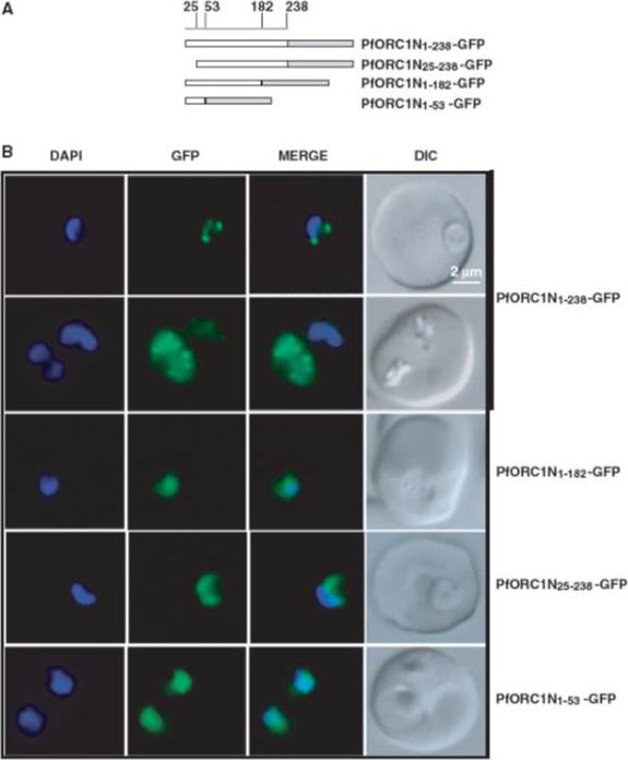
Fluorescence microscopy of different N-terminal domains as GFP-fusion proteins. (A) Schematic diagrams of different N-terminal domains fused with GFP as indicated in the figure. The numbers on the top indicate the positions of the amino acid residues. (B) Live cell imaging of different parasite lines as indicated on the right. ORC1N1–238–GFP predominantly shows punctate staining at the nuclear periphery either at the ring stage (first row) or at the later stage (second row). None of the other constructs show nuclear punctate staining pattern. ORC1N25–238–GFP shows diffused staining pattern all over the parasite while both ORC1N1–182–GFP and ORC1N1–53–GFP shows enrichment of GFP signal in the nucleus. ORC1N1–238 binds to DNA and forms nuclear periphery punctate foci whereas ORC1N1–182 fails to do so suggesting that residues between 182 and 238 are involved in DNA binding.Deshmukh AS, Srivastava S, Herrmann S, Gupta A, Mitra P, Gilberger TW, Dhar SK. The role of N-terminus of Plasmodium falciparum ORC1 in telomeric localization and var gene silencing. Nucleic Acids Res. 2012 40(12):5313-31
See original on MMP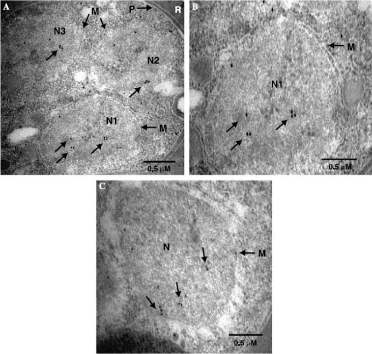
The secondary antibodies were conjugated to 60–80 nm gold particles. (A) Parasite-infected RBC probed with anti-PfORC1 antibodies. Three nuclei (N1, N2, and N3) are shown in the frame. Arrowheads indicate the deposition of the gold particles within individual nucleus. M - nuclear membrane and P - parasite membrane. R - RBC. The scale bar is shown at the bottom right. (B) A higher resolution magnified version of N1 from (A). Positions of the gold particle deposition and the nuclear membrane (M) are shown. (C) The pattern of the PfORC1 distribution in an individual nucleus of a parasite-infected RBC (different from A). Mehra P, Biswas AK, Gupta A, Gourinath S, Chitnis CE, Dhar SK. Expression and characterization of human malaria parasite Plasmodium falciparum origin recognition complex subunit 1. Biochem Biophys Res Commun. 2005 337:955-66. Copyright Elsevier 2010.
See original on MMP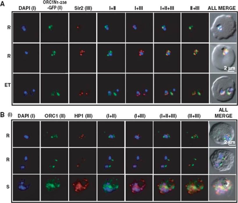
Co-localization of ORC1N1–238–GFP and Sir2 and immunolocalization of ORC1 and HP1 in 3D7. Glass slides containing the parasite smears from ORC1N1–238–GFP expressing parasites or 3D7 wild-type parasite lines were treated for immunolocalization using respective antibodies as indicated in the panels (A) and (B). (A) Co-localization of GFP and Sir2 in ORC1N1–238–GFP parasites during early stages of development as indicated on the left. ‘R’ indicates ring stage and ‘ET’ indicates early trophozoite stage parasites. DAPI shows the nuclei. Both ORC1 and Sir2 are localized to the nuclear periphery during the ring or early trophozoite stage. However, both proteins reorganize themselves at the onset of DNA replication and spread in the nucleus and cytoplasm. B(i): Both ORC1 and HP1 show nuclear punctate staining in 3D7 wild-type parasites during the early stages of development. ‘S’ indicates schizont stage parasites.Deshmukh AS, Srivastava S, Herrmann S, Gupta A, Mitra P, Gilberger TW, Dhar SK. The role of N-terminus of Plasmodium falciparum ORC1 in telomeric localization and var gene silencing. Nucleic Acids Res. 2012 40(12):5313-31
See original on MMP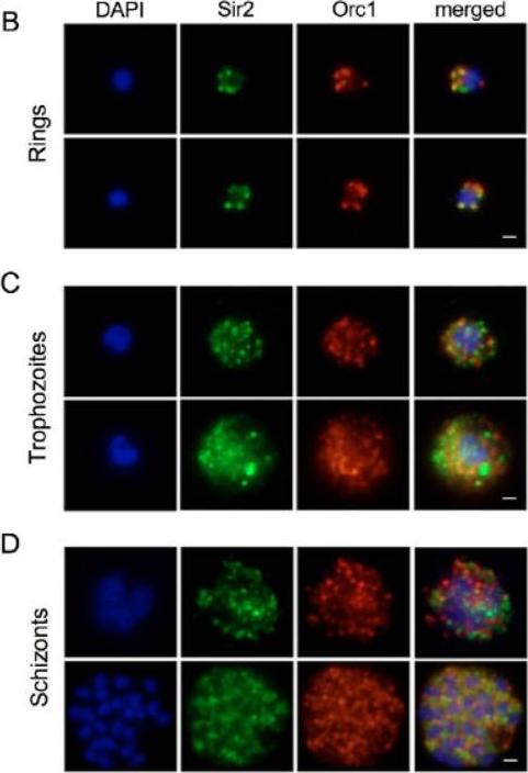
Sir2 and Orc1 relocalize during the developmental cycle of P. falciparum. (B-D) IF analysis of Sir2 (green) and Orc1 (red) during the blood-stage cycle. (B) Ring stages display a punctate pattern at the nuclear periphery. (C) In the trophozoite stages, anti-Sir2 and -Orc1 antibodies reveal an apparent increase of both protein levels, and an additional punctate and diffuse pattern inside and outside of the nucleus. (D) The schizont stage, Sir2 and Orc1 seem to relocalize at the nuclear periphery.Mancio-Silva L, Rojas-Meza AP, Vargas M, Scherf A, Hernandez-Rivas R. Differential association of Orc1 and Sir2 proteins to telomeric domains in Plasmodium falciparum. J Cell Sci. 2008 121:2046-53.
See original on MMP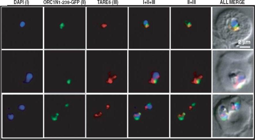
Fluorescence in situ hybridization coupled immunofluorescence assay (FISH–IFA). (FISH–IFA was performed using fluorescence analog labeled TARE-6 DNA probe and antibodies against GFP in ORC1N1–238–GFP expressing parasites. The results indicate that majority of the TARE-6 and ORC1N1–238–GFP signals are overlapping or closely associated with each other. Top panel corresponds to ring stage parasite and bottom two panels correspond to trophozoite stage parasites.Deshmukh AS, Srivastava S, Herrmann S, Gupta A, Mitra P, Gilberger TW, Dhar SK. The role of N-terminus of Plasmodium falciparum ORC1 in telomeric localization and var gene silencing. Nucleic Acids Res. 2012 40(12):5313-31
See original on MMP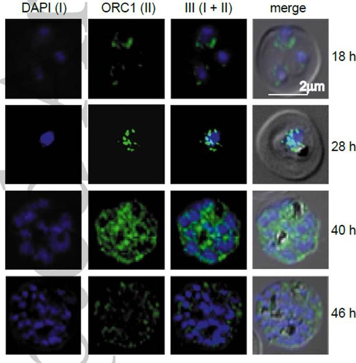
Endogenous ORC1 is redistributed during blood-stage asexual cycle. Immunofluorescence analysis using antibodies against PfORC1 reveals punctate pattern at the nuclear periphery in the ring stage (~18 hr) and early-to-mid trophozoite stage (~28 hr). At a later stage (~40 hr), an apparent increase of protein level and diffused pattern in the cytoplasm is observed, whereas during very late schizont (~46 hr), the PfORC1 signal decreases drastically.Deshmukh AS, Agarwal M, Mehra P, Gupta A, Gupta N, Doerig CD, Dhar SK. Regulation of Plasmodium falciparum Origin Recognition Complex subunit 1 (PfORC1) function through phosphorylation mediated by CDK like kinase PK5. Mol Microbiol. 2015 [Epub ahead of print]
See original on MMP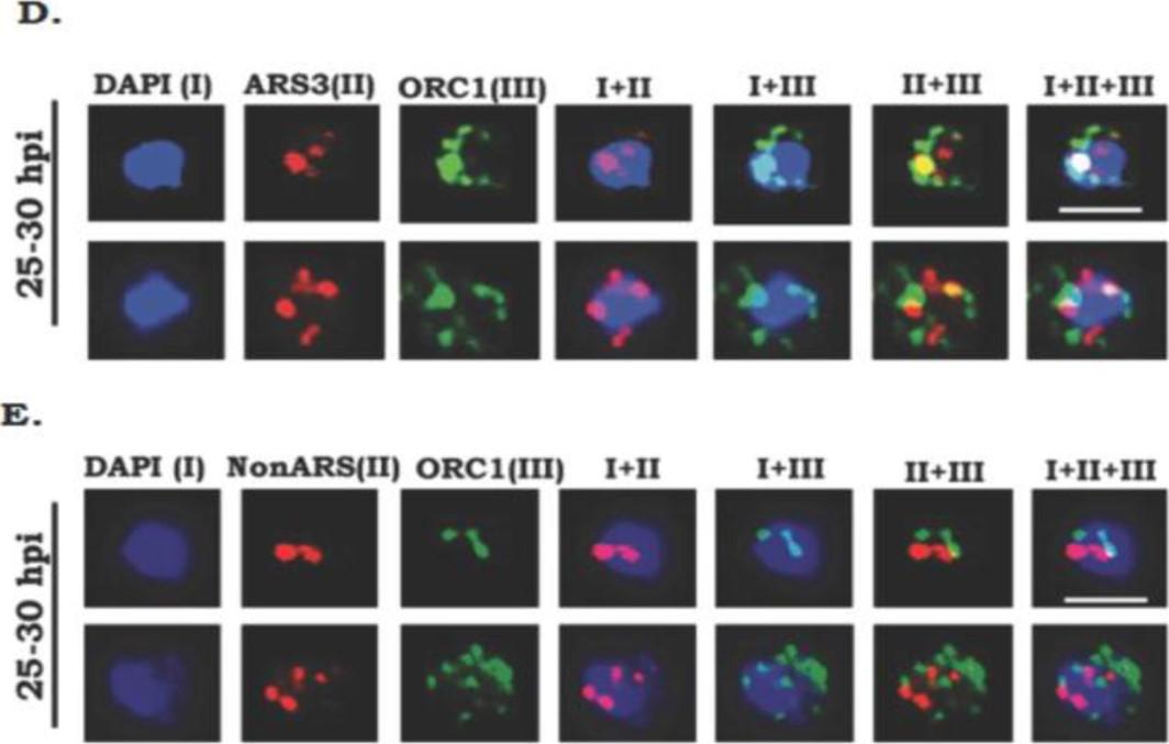
Endogenous PfORC1 binds to PfARS sequences in Plasmodium falciparum. FISH-IFA was performed using fluorescence labeled ARS3 DNA probe and antibodies against ORC1 in the trophozoite stage (25-30hpi) parasites. Results indicate ~75% co-localization of PfARS3 signals with ORC1.Right panel shows the graphical representation of merged signals versus apart signals. (E) FISH-IFA experiments were performed using non-ARS sequence as a probe. Co-localization of non-ARS signals with ORC1was rare (<10%). Right panel shows the graphical representation of co-localized signals versus non-colocalized signals for twenty images in each case. Agarwal M, Bhowmick K, Shah K, Krishnamachari A, Dhar SK. Identification and Characterization of ARS like sequences as putative origin(s) of replication in human malaria parasite Plasmodium falciparum. FEBS J. 2017 Jun 23. [Epub ahead of print]
See original on MMP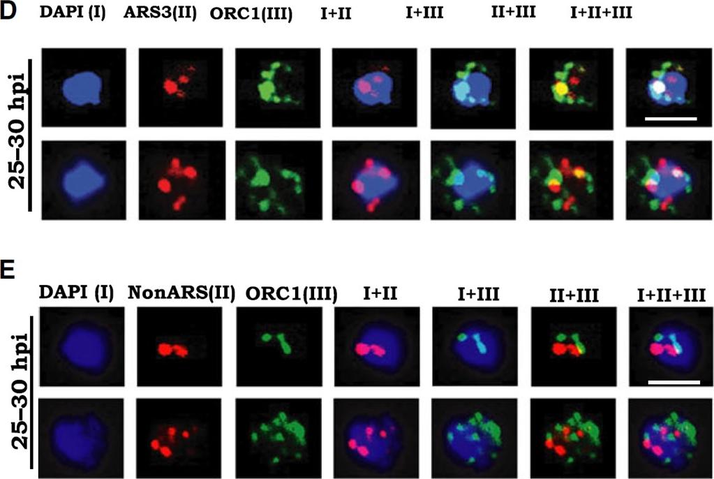
Endogenous PfORC1 binds to PfARS sequences in Plasmodium falciparum. FISH-IFA was performed using fluorescence labeled ARS3 DNA probe and antibodies against ORC1 in the trophozoite stage (25–30 hpi) parasites. Results indicate ~ 75% colocalization of PfARS3 signals with ORC1. Right panel shows the graphical representation of merged signals versus separate signals. (E) FISH-IFA experiments were performed using non-ARS sequence as a probe. Colocalization of non-ARS signals with ORC1 was rare (< 10%). Right panel shows the graphical representation of colocalized signals versus non-colocalized signals for 20 images in each case. The scale bar is 2 mm.Agarwal M, Bhowmick K, Shah K, Krishnamachari A, Dhar SK. Identification and characterization of ARS-like sequences as putative origin(s) of replication in human malaria parasite Plasmodium falciparum. FEBS J. 2017 Aug;284(16):2674-2695.
See original on MMPMore information
| PlasmoDB | PBANKA_0602000 |
| GeneDB | PBANKA_0602000 |
| Malaria Metabolic Pathways | Localisation images Pathways mapped to |
| Previous ID(s) | PB000867.02.0, PB401668.00.0, PBANKA_060200 |
| Orthologs | PCHAS_0603800 , PF3D7_1203000 , PKNH_1302900 , PVP01_1302300 , PVX_084195 , PY17X_0604500 |
| Google Scholar | Search for all mentions of this gene |