PBANKA_0515200 MORN repeat-containing protein 1, putative (MORN1)
Mutant phenotypes [+]
| Species | Stage | Phenotype | Reference | Submitter |
|---|---|---|---|---|
| P. berghei ANKA | Asexual |
No difference |
PlasmoGEM (Barseq) | PlasmoGEM |
| P. falciparum 3D7 | Asexual |
No difference |
34878291 \"In this work, we demonstrate that the P. falciparum membrane occupation and recognition nexus repeat-containing protein 1 (PfMORN1) is not required for asexual replication. Following inducible knockout of PfMORN1, we find no detectable defect in asexual parasite morphology or replicative fitness.\" |
Theo Sanderson, Francis Crick Institute |
Imaging data (from Malaria Metabolic Pathways)
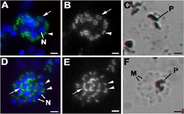
MORN1 in schizogony of Plasmodium. Images of infected erythrocytes immunostained with anti-MORN1 (green) and the nuclei counterstain showing the DAPI-positive nucleus (blue) (A and D). Panels B and E present the green channel in gray, increasing the clarity of the MORN1-positive staining. Panels C and F represent the transmitted light image. (A to C) Infected erythrocyte illustrating a late stage in schizogony in which 16 individual MORN1-positive rings (tangential appearance [arrows]) or plaques (longitudinal orientation [arrowheads]), representing the free ends of the cone-shaped IMCs, can be identified. (D to F) Infected erythrocyte containing a mature schizont showing the DAPI staining of nuclei (N) of the daughters with strong posterior MORN1 staining (arrows) and a smaller, more apical MORN1-positive structure, possibly representing the nuclear pole (arrowheads). The transmitted light images illustrate the dark-appearing hemozoin pigment (P) in panels C and F. In panel F, individual merozoites (M) can be identified. Bars = 1 mm. MORN1 localizes to the merozoite budding sites on Plasmodium schizonts.Ferguson DJ, Sahoo N, Pinches RA, Bumstead JM, Tomley FM, Gubbels MJ. MORN1 has a conserved role in asexual and sexual development across the apicomplexa. Eukaryot Cell. 2008 7:698-711.
See original on MMP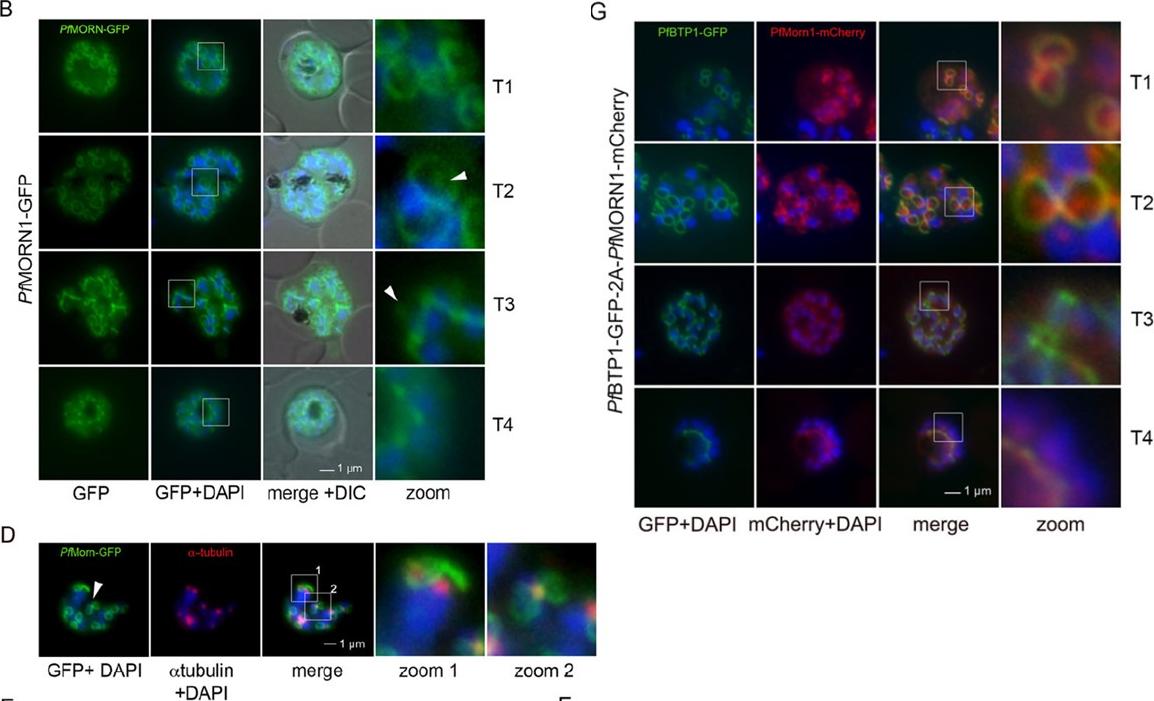
Colocalization of BTP1 with MORN1. (B) Localization of P. falciparum (pf)MORN1–GFP in unfixed parasites. MORN1–GFP reveals a contractile ring structure that moves along the longitudinal axes of the nascent merozoite (T1–T4) with an additional punctate structure that might represent the centrocone (marked with arrowheads). Nuclei were stained with DAPI (blue). Enlargement of selected areas are marked with a white square and referred to as ‘zoom’ (a fourfold magnification). (D) Colocalization of MORN1–GFP with α-tubulin (red) that highlights the microtubule-organizing centers (MTOCs) at this stage. Although the MORN1-marked ring structure is clearly distinct from the MTOC (zoom 1), there is a close association of the MTOC with the additional MORN1-marked structure (zoom 2). (G) Colocalization analysis of BTP1–GFP (green) with MORN1–mCherry (red) revealed their identical localization during schizogony, except for the exclusively MORN1–mCherry-highlighted putative centrocone.Kono M, Heincke D, Wilcke L, Wong TW, Bruns C, Herrmann S, Spielmann T, Gilberger TW. Pellicle formation in the malaria parasite. J Cell Sci. 2016 129(4):673-80. PMID: 26763910
See original on MMPMore information
| PlasmoDB | PBANKA_0515200 |
| GeneDB | PBANKA_0515200 |
| Malaria Metabolic Pathways | Localisation images Pathways mapped to |
| Previous ID(s) | PB000603.00.0, PB300190.00.0, PBANKA_051520 |
| Orthologs | PCHAS_0515300 , PF3D7_1031200 , PKNH_0615900 , PVP01_0616300 , PVX_111165 , PY17X_0516200 |
| Google Scholar | Search for all mentions of this gene |