PBANKA_0512800 merozoite TRAP-like protein, putative (MTRAP)
Disruptability [+]
| Species | Disruptability | Reference | Submitter | |
|---|---|---|---|---|
| P. berghei ANKA |
Possible |
27832590 Gametes fail to egress |
Theo Sanderson, WTSI | |
| P. berghei ANKA |
Possible |
RMgm-4056 | Imported from RMgmDB | |
| P. berghei ANKA |
Possible |
RMgm-1493 | Imported from RMgmDB | |
| P. berghei ANKA |
Possible |
RMgm-4519 | Imported from RMgmDB | |
| P. falciparum 3D7 |
Refractory |
16321976 | Theo Sanderson, Wellcome Trust Sanger Institute | |
| P. falciparum 3D7 |
Possible |
27832590 Gametes fail to egress |
Theo Sanderson, WTSI | |
| P. falciparum 3D7 |
Possible |
USF piggyBac screen (Insert. mut.) | USF PiggyBac Screen | |
Mutant phenotypes [+]
| Species | Stage | Phenotype | Reference | Submitter |
|---|---|---|---|---|
| P. berghei ANKA | Asexual |
No difference |
RMgm-4056 | Imported from RMgmDB |
| P. berghei ANKA | Asexual |
No difference |
RMgm-4519 | Imported from RMgmDB |
| P. berghei ANKA | Gametocyte |
Attenuated |
27832590 Gametes fail to egress |
Theo Sanderson, WTSI |
| P. berghei ANKA | Gametocyte |
Difference from wild-type |
RMgm-4056
Normal numbers of gametocytes are produced. Both male and female gametes cannot escape from the host red blood cell after activation of gametocytes. Evidence is presented that the parasitophorous membrane is not disrupted in the absence of MTRAP |
Imported from RMgmDB |
| P. berghei ANKA | Gametocyte |
Difference from wild-type |
RMgm-1493
Normal numbers of gametocytes are produced. Male and female gametocytes show a defect in egress from the host erythrocyte. |
Imported from RMgmDB |
| P. berghei ANKA | Gametocyte |
No difference |
RMgm-4519 | Imported from RMgmDB |
| P. berghei ANKA | Ookinete |
Difference from wild-type |
RMgm-4056
No fertilisation and ookinetes. Normal numbers of gametocytes are produced. Both male and female gametes can escape from the hosr red blood cell after activation of gametocytes. Evidence is presented that the parasitophorous membrane is not disrupted in the absence of MTRAP |
Imported from RMgmDB |
| P. berghei ANKA | Ookinete |
Difference from wild-type |
RMgm-1493
Male and female gametocytes show a defect in egress from the host erythrocyte. Mutant males gametes are trapped within the RBC yet motile. No ookinetes are formed in vitro |
Imported from RMgmDB |
| P. berghei ANKA | Oocyst |
Difference from wild-type |
RMgm-4056
No oocyst production |
Imported from RMgmDB |
| P. berghei ANKA | Oocyst |
Difference from wild-type |
RMgm-1493
No oocyst production in A. stephensi mosquitoes |
Imported from RMgmDB |
| P. falciparum 3D7 | Gametocyte |
Attenuated |
27832590 Gametes fail to egress |
Theo Sanderson, WTSI |
Imaging data (from Malaria Metabolic Pathways)
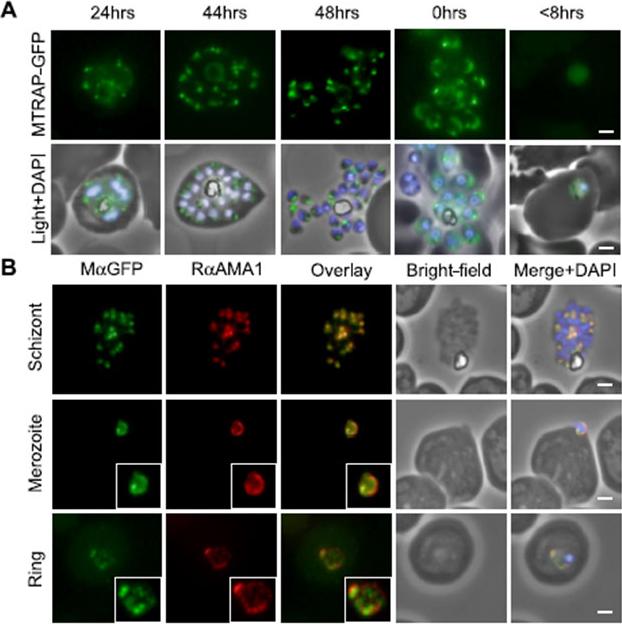
MTRAP undergoes extensive processing during invasion. A, episomally expressed MTRAP-GFP followed through the life cycle confirms apical localization (44–48 h) suggests the release of MTRAP onto the surface of free merozoites (capping, 0 h) and suggests that, despite processing, the cytoplasmic tail is carried through to newly invaded rings (<8 h). B, this is confirmed by following co-localization with antibodies that recognize AMA1, which have been previously reported as being carried through to rings. In early ring stages, there does not appear to be direct co-localization, suggesting that they may be associating with different compartments. Baum J, Richard D, Healer J, Rug M, Krnajski Z, Gilberger TW, Green JL, Holder AA, Cowman AF. A conserved molecular motor drives cell invasion and gliding motility across malaria life cycle stages and other apicomplexan parasites. J Biol Chem. 2006 281(8):5197-208.
See original on MMP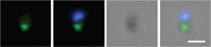
Schizont rich culture was thin smeared on a glass slide, fixed in methanol, stainined with rabbit anti-MTRAP polyclonal serum (1:200) and secondary Alexa-Flor488 anti-Rabbit. Dual colour fluorescence images were captured using a Carl Zeiss Axioskop 2 microscope at 100x using Oil imersion. Nucleus is marked with DAPI. Scale bar indicate 1µM. Free merozoite is shown with MTRAP clearly marking the apical end (shown to be micronemes by immuno-EM).Baum J, Richard D, Healer J, Rug M, Krnajski Z, Gilberger TW, Green JL, Holder AA, Cowman AF. A conserved molecular motor drives cell invasion and gliding motility across malaria life cycle stages and other apicomplexan parasites. J Biol Chem. 2006 281(8):5197-208.
See original on MMP
Confocal fluorescence microscopy images of transfected 3D7 P. falciparum-infected RBCs expressing a GFP chimera directed to the micronemes. Phase image, the GFP fluorescence signal and an overlay of a P. falciparum transfectant expressing the merozoite thrombospondin-related anonymous protein. Scale bar = 5 µmTilley L, McFadden G, Cowman A, Klonis N. Illuminating Plasmodium falciparum-infected red blood cells. Trends Parasitol. 2007 23:268-77
See original on MMPMTRAP is cleaved within the micronemes. A) IFA of compound 1 (C1) treated schizonts co-labelled with mAMA1 antibodies (green) and rM-tsr (top panels), rM-mid (middle panels) or rM-tail (bottom panels) (red in respective images). Scale bars = 1 μm. C) Schematic of pre- (left half) and post- (right half) invasion merozoites, showing the predominant distribution of each protein tested as part of this study. It should be noted that all proteins show a degree of variability in labelling distributions across merozoites imaged to an extent that is not possible to depict here.Riglar DT, Whitehead L, Cowman AF, Rogers KL, Baum J. Localization-based imaging of malarial antigens during red cell entry reaffirms role for AMA1 but not MTRAP in invasion. J Cell Sci. 2015. [Epub ahead of print]
See original on MMP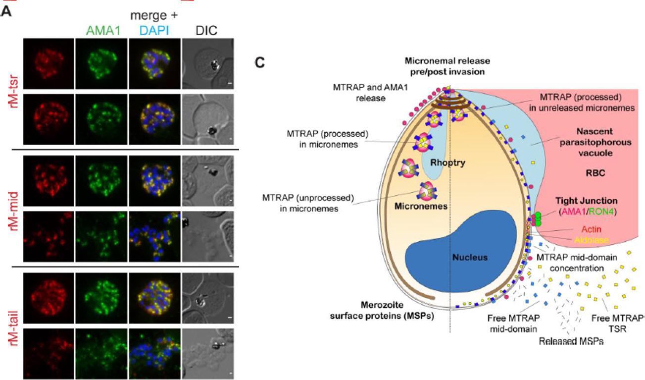
MTRAP is cleaved within the micronemes. A) IFA of compound 1 (C1) treated schizonts co-labelled with mAMA1 antibodies (green) and rM-tsr (top panels), rM-mid (middle panels) or rM-tail (bottom panels) (red in respective images). Scale bars = 1 μm. C) Schematic of pre- (left half) and post- (right half) invasion merozoites, showing the predominant distribution of each protein tested as part of this study. It should be noted that all proteins show a degree of variability in labelling distributions across merozoites imaged to an extent that is not possible to depict here.Riglar DT, Whitehead L, Cowman AF, Rogers KL, Baum J. Localization-based imaging of malarial antigens during red cell entry reaffirms role for AMA1 but not MTRAP in invasion. J Cell Sci. 2015. [Epub ahead of print]
See original on MMP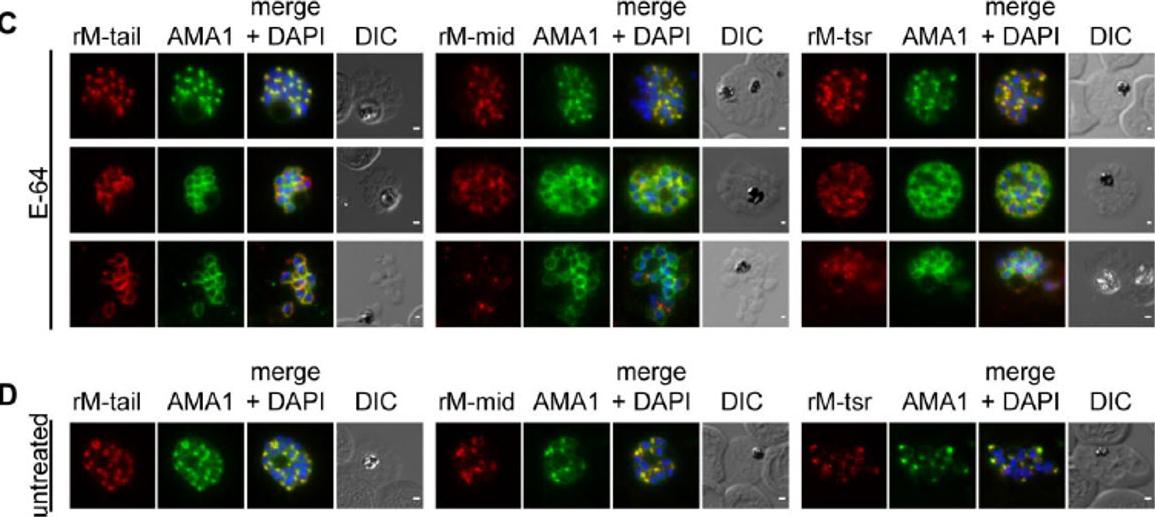
MTRAP is cleaved irrespective of both invasion and egress from the schizont. (C) treated with E-64 to prevent egress or (D) from untreated culture, co-labelled AMA1 monoclonal antibodies (green) and rM-tail (left), rM-mid (middle) or rM-tsr (right) (red in respective images). Scale bars = 1 μm.Riglar DT, Whitehead L, Cowman AF, Rogers KL, Baum J. Localization-based imaging of malarial antigens during red cell entry reaffirms role for AMA1 but not MTRAP in invasion. J Cell Sci. 2015. [Epub ahead of print]
See original on MMP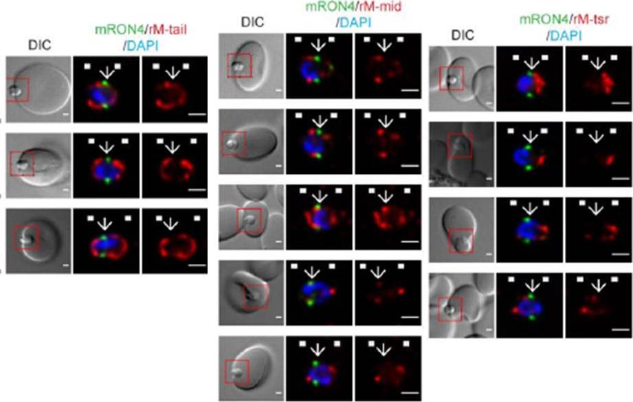
Longitudinal intensity profiling identifies unexpected localisation profiles for MTRAP during merozoite invasion. A-C). Single-slice images of example merozoites of mRON4 (green) vs (Left panle) rMTRAP-tail (rM-tail) (red), (midle panel) rMTRAP mid (rM-mid) (red), or (right panel) rMTRAP tsr (rM-tsr). Scale bars = 1 μm. Red boxes in DIC images indicate zoomed regions for middle and right panels. Arrows indicate the position of the RON4 labelled tight junction.Riglar DT, Whitehead L, Cowman AF, Rogers KL, Baum J. Localization-based imaging of malarial antigens during red cell entry reaffirms role for AMA1 but not MTRAP in invasion. J Cell Sci. 2015. [Epub ahead of print]
See original on MMP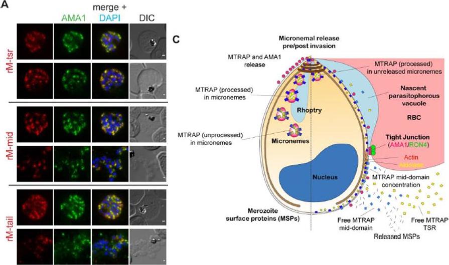
MTRAP is cleaved within the micronemes. A) IFA of compound 1 (C1) treated schizonts co-labelled with mAMA1 antibodies (green) and rM-tsr (top panels), rM-mid (middle panels) or rM-tail (bottom panels) (red in respective images). Scale bars = 1 μm. C) Schematic of pre- (left half) and post- (right half) invasion merozoites, showing the predominant distribution of each protein tested as part of this study. It should be noted that all proteins show a degree of variability in labelling distributions across merozoites imaged to an extent that is not possible to depict here.Riglar DT, Whitehead L, Cowman AF, Rogers KL, Baum J. Localization-based imaging of malarial antigens during red cell entry reaffirms role for AMA1 but not MTRAP in invasion. J Cell Sci. 2015. [Epub ahead of print]
See original on MMP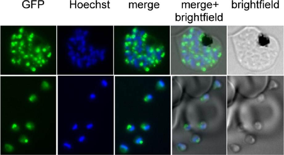
Expression of GFP-tagged MTRAP in asexual blood stage parasites. Fluorescent images of live parasites expressing GFP-tagged MTRAP (green); nuclei (blue) were stained with Hoechst dye prior to microscopy. Merged and bright-field images are also shown. The GFPtagged MTRAP is seen in a subcellular location suggestive of an apical organelle. Diaz SA, Martin SR, Howell SA, Grainger M, Moon RW, Green JL, Holder AA. The Binding of Plasmodium falciparum Adhesins and Erythrocyte Invasion Proteins to Aldolase Is Enhanced by Phosphorylation. PLoS One. 2016 Sep 8;11(9):e0161850.
See original on MMPMore information
| PlasmoDB | PBANKA_0512800 |
| GeneDB | PBANKA_0512800 |
| Malaria Metabolic Pathways | Localisation images Pathways mapped to |
| Previous ID(s) | PB000355.03.0, PBANKA_051280 |
| Orthologs | PCHAS_0512900 , PF3D7_1028700 , PKNH_0613400 , PVP01_0613800 , PVX_111290 , PY17X_0513900 |
| Google Scholar | Search for all mentions of this gene |