PBANKA_0505100 ADP-ribosylation factor, putative (ARF1)
Disruptability [+]
| Species | Disruptability | Reference | Submitter | |
|---|---|---|---|---|
| P. berghei ANKA |
Refractory |
PlasmoGEM (Barseq) | PlasmoGEM | |
| P. falciparum 3D7 |
Refractory |
USF piggyBac screen (Insert. mut.) | USF PiggyBac Screen | |
Mutant phenotypes [+]
None reported yet. Please press the '+' button above to add one.Imaging data (from Malaria Metabolic Pathways)
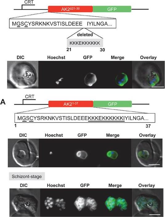
Upper panel: Deletion of the stretch of basic residues at the N-terminus of PfAK2 alters the subcellular location of the protein. (A) The AK2Δ21-30/GFP was expressed using the CRT promoter (construct indicated above the images). Live cell imaging of the PfAK2Δ21-30/GFP parasite line in a late trophozoite stage parasite.Lower panel: The PfAK2 N-terminus targets GFP to the outside of the parasite plasma membrane. (A) The AK21-37/GFP was expressed using the CRTpromoter (construct indicated above the images). Live cell imaging of the PfAK21-37/GFP in trophozoite and schizont stages, showed a location of the GFP signal similar to that of PfAK2/GFP. In the schizont stage of these parasites a clear signal for the fusion protein was visible around each of the individualdaughter merozoites.Thavayogarajah T, Gangopadhyay P, Rahlfs S, Becker K, Lingelbach K, Przyborski JM, Holder AA. Alternative Protein Secretion in the Malaria Parasite Plasmodium falciparum. PLoS One. 2015 Apr 24;10(4):e0125191.
See original on MMP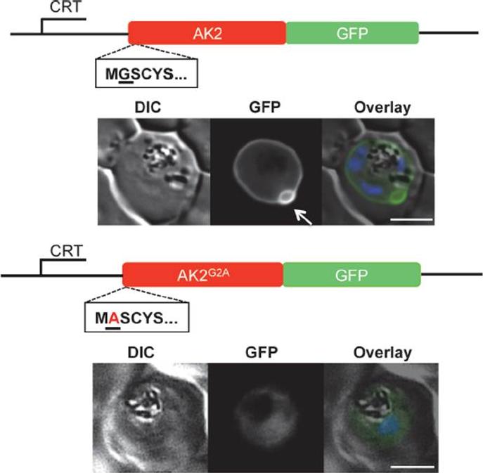
Subcellular location of PfAK2/GFP and the non-myristoylated G2A variant, as determined by live fluorescence microscopy (A) The AK2/GFP and the AK2G2A/GFP fusion proteins were expressed using the CRT promoter from an episomal plasmid (constructs indicated above the images). AK2/GFP is located at the periphery of the intracellular parasite as judged by epifluorescence microscopy, and is associated with one or two protuberances towards the host cell cytoplasm present on each parasite (indicated by white arrow). In contrast, the AK2G2A/GFP chimera is located within the parasite cytosol. The infected cell was visualised by differential interference contrast (DIC), intrinsic fluorescence of the GFP identified the location of the AK2/GFP fusion protein, and parasite nuclei were detected by Hoechst staining. Overlay: green (GFP), blue (DNA). Scale bar-3 μm.Thavayogarajah T, Gangopadhyay P, Rahlfs S, Becker K, Lingelbach K, Przyborski JM, Holder AA. Alternative Protein Secretion in the Malaria Parasite Plasmodium falciparum. PLoS One. 2015 Apr 24;10(4):e0125191.
See original on MMPMore information
| PlasmoDB | PBANKA_0505100 |
| GeneDB | PBANKA_0505100 |
| Malaria Metabolic Pathways | Localisation images Pathways mapped to |
| Previous ID(s) | PB000446.01.0, PBANKA_050510 |
| Orthologs | PCHAS_0505200 , PF3D7_1020900 , PKNH_0605200 , PVP01_0606100 , PVX_001905 , PY17X_0506200 |
| Google Scholar | Search for all mentions of this gene |