PBANKA_0416700 bacterial histone-like protein, putative (HU)
Disruptability [+]
| Species | Disruptability | Reference | Submitter | |
|---|---|---|---|---|
| P. berghei ANKA |
Refractory |
RMgm-266 | Imported from RMgmDB | |
| P. falciparum 3D7 |
Refractory |
USF piggyBac screen (Insert. mut.) | USF PiggyBac Screen | |
Mutant phenotypes [+]
None reported yet. Please press the '+' button above to add one.Imaging data (from Malaria Metabolic Pathways)
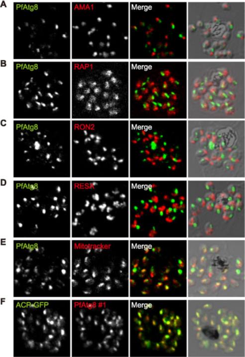
PfAtg8 localizes to the apicoplast. P. falciparum FCR3 (A–E) and P. falciparum 3D7 transfected with ACP-GFP (F–H) were stained with the indicated organelle markers and visualized by confocal microscopy (because ACP-GFP was not uniformly expressed, some merozoites displayed only faint GFP signals). Anti-PfAtg8 antibody #1 was used in (A–F), and anti-PfAtg8 antibody #2 was used in (G). Apical membrane antigen 1 (AMA1) as a microneme marker (A), rhoptry-associated protein 1 (RAP1) as a rhoptry body marker (B), rhoptry neck protein 2 (RON2) as a rhoptry neck marker (C), the ring-infected erythrocyte surface antigen (RESA) as a dense granule marker (D), MitoTrackerRed CMXRos as a mitochondria marker (E), ACPGFP (F–H) and the organellar histone-like protein PfHU (H) as an apicoplast marker were used. Scale bar, 1 mm.Kitamura K, Kishi-Itakura C, Tsuboi T, Sato S, Kita K, Ohta N, Mizushima N. Autophagy-Related Atg8 Localizes to the Apicoplast of the Human Malaria Parasite Plasmodium falciparum. PLoS One. 2012;7(8):e42977.
See original on MMP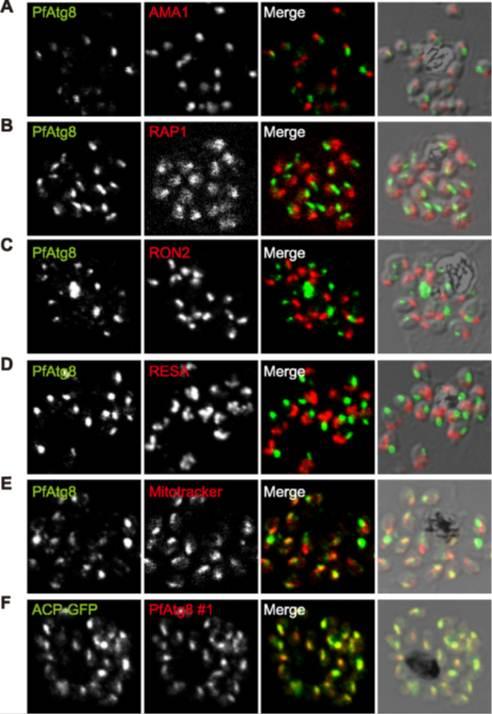
PfAtg8 localizes to the apicoplast. P. falciparum FCR3 (A–E) and P. falciparum 3D7 transfected with ACP-GFP (F–H) were stained with the indicated organelle markers and visualized by confocal microscopy (because ACP-GFP was not uniformly expressed, some merozoites displayed only faint GFP signals). Anti-PfAtg8 antibody #1 was used in (A–F), and anti-PfAtg8 antibody #2 was used in (G). Apical membrane antigen 1 (AMA1) as a microneme marker (A), rhoptry-associated protein 1 (RAP1) as a rhoptry body marker (B), rhoptry neck protein 2 (RON2) as a rhoptry neck marker (C), the ring-infected erythrocyte surface antigen (RESA) as a dense granule marker (D), MitoTrackerRed CMXRos as a mitochondria marker (E), ACPGFP (F–H) and the organellar histone-like protein PfHU (H) as an apicoplast marker were used. Scale bar, 1 mm.Kitamura K, Kishi-Itakura C, Tsuboi T, Sato S, Kita K, Ohta N, Mizushima N. Autophagy-Related Atg8 Localizes to the Apicoplast of the Human Malaria Parasite Plasmodium falciparum. PLoS One. 2012;7(8):e42977
See original on MMP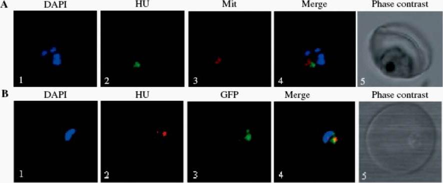
Localization of PfHU. (A) Immunolocalization of PfHU using confocal microscopy. Panels show nuclear DNA staining with DAPI (1), PfHU fluorescence (2), MitoTracker Red signal (3) and their overlap (4) in a late trophozoite. The corresponding phase–contrast scan is shown in (5). (B) Co-localization of PfHU and apicoplast-targeted GFP. Nuclear DNA stained with DAPI (1), PfHU signal (2), GFP signal (3) and their overlay (4) are shown. PfHU is associated with apicoplast DNA and is involved in DNA compaction.Ram EV, Naik R, Ganguli M, Habib S. DNA organization by the apicoplast-targeted bacterial histone-like protein of Plasmodium falciparum. Nucleic Acids Res. 2008 36(15):5061-73.
See original on MMP
Immunofluorescence localisation of PfSufS and PfSufE. (A) Immunofluorescence assay of P. falciparum 3D7 cells using anti-PfSufS Ab with anti-PfHU Ab as apicoplast marker (panel i) or Mitotracker Red (panel ii) indicates targeting of PfSufS to the apicoplast. (B) Co-localisation of PfSufE and apicoplast-targeted GFP observed in the P. falciparum D10 ACPleader-GFP cell line using anti-PfSufE and anti-GFP antibodies (panel i). No overlap of PfSufE signal was observed with Mitotracker Red in P. falciparum 3D7 cells (panel ii) indicating apicoplast localization of PfSufE. PfSufS co-localised with PfHU, an apicoplast DNA condensation protein and no overlapping signals for PfSufS were seen with the mitochondrial marker Mitotracker Red in P. falciparum 3D7 cells (A). PfSufE was localised in the P. falciparum D10-ACPleader-GFP line that targets GFP to the apicoplast. Specific PfSufE signals overlapping with apicoplast-targeted GFP were observed and no overlap of PfSufE signal was seen with Mitotracker Red (B). The localisation of PfSufS and PfSufE exclusively to the apicoplast was thus confirmed.Charan M, Singh N, Kumar B, Srivastava K, Siddiqi MI, Habib S. Sulphur mobilisation for [Fe-S] cluster assembly by the essential SUF pathway in the Plasmodium falciparum apicoplast and its inhibition. Antimicrob Agents Chemother. 2014 Apr 7. [Epub ahead of print]
See original on MMP
Immunofluorescence localisation of PfSufS and PfSufE. (A) Immunofluorescence assay of P. falciparum 3D7 cells using anti-PfSufS Ab with anti-PfHU Ab as apicoplast marker (panel i) or Mitotracker Red (panel ii) indicates targeting of PfSufS to the apicoplast. (B) Co-localisation of PfSufE and apicoplast-targeted GFP observed in the P. falciparum D10 ACPleader-GFP cell line using anti-PfSufE and anti-GFP antibodies (panel i). No overlap of PfSufE signal was observed with Mitotracker Red in P. falciparum 3D7 cells (panel ii) indicating apicoplast localization of PfSufE. PfSufS co-localised with PfHU, an apicoplast DNA condensation protein and no overlapping signals for PfSufS were seen with the mitochondrial marker Mitotracker Red in P. falciparum 3D7 cells (A). PfSufE was localised in the P. falciparum D10-ACPleader-GFP line that targets GFP to the apicoplast. Specific PfSufE signals overlapping with apicoplast-targeted GFP were observed and no overlap of PfSufE signal was seen with Mitotracker Red (B). The localisation of PfSufS and PfSufE exclusively to the apicoplast was thus confirmed.Charan M, Singh N, Kumar B, Srivastava K, Siddiqi MI, Habib S. Sulphur mobilisation for [Fe-S] cluster assembly by the essential SUF pathway in the Plasmodium falciparum apicoplast and its inhibition. Antimicrob Agents Chemother. 2014 Apr 7. [Epub ahead of print
See original on MMP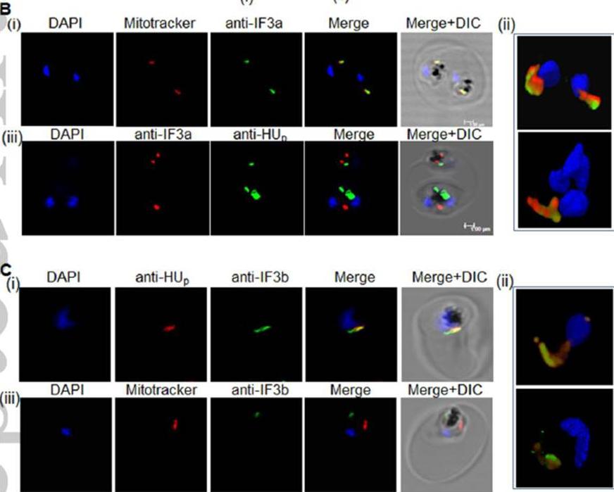
Immunofluorescence localization of PfIF3a and PfIF3b. (B) Panel i, P. falciparum-infected erythrocytes stained with Mitotracker Red and probed with PfIF3a antiserum; panel ii, 3-dimensional reconstruction after z-sectioning shows clear overlay of the PfIF3a signal with the mitochondrion; panel iii, Absence of overlap of the PfIF3a signal with apicoplast PfHU. (C) Panel i, parasitized erythrocytes probed with anti-IF3b and anti-HUp anti-sera; panel ii, 3-dimensional reconstruction shows overlap of IF3b with apicoplast HUp; panel iii, PfIF3b signal does not overlap with Mitotracker Red. Nuclear DNA is stained with DAPI.Haider A, Allen SM, Jackson KE, Ralph SA, Habib S. Targeting and function of proteins mediating translation initiation in organelles of Plasmodium falciparum. Mol Microbiol. 2015 Feb 17. [Epub ahead of print]
See original on MMP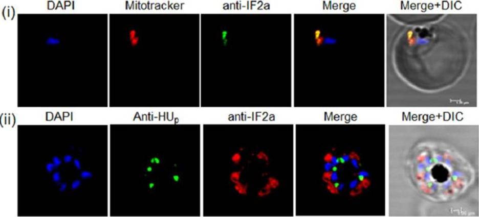
Immunofluorescence assay (IFA) with anti-PfIF2a-C serum and the mitochondrial dye Mitotracker Red CMXRos (panel i); IFA to detect the apicoplast marker protein PfHU (using PfHUp antiserum) and PfIF2a (panel ii). Parasite nuclear DNA is stained with DAPI.Haider A, Allen SM, Jackson KE, Ralph SA, Habib S. Targeting and function of proteins mediating translation initiation in organelles of Plasmodium falciparum. Mol Microbiol. 2015 Feb 17. [Epub ahead of print]
See original on MMP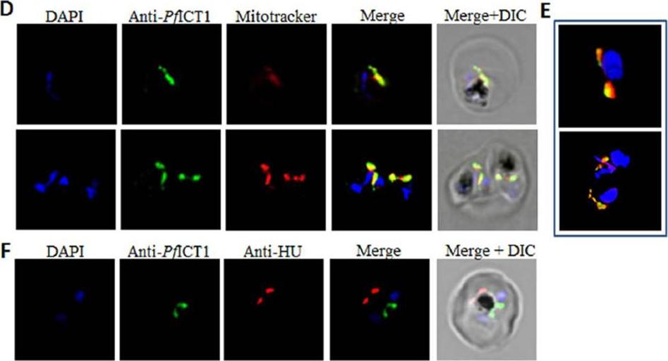
Molecular modeling, detection and subcellular localization of PfICT1. (D-F) Immunofluorescence assay for localization of PfICT1 using purified anti-PfICT1 Ab. Overlap of the PfICT1 signal was seen with the mitochondrial dye (D) and 3-dimensional reconstruction after z-sectioning showed clear overlay of PfICT1 signal with the mitochondrion (E). No overlap was observed with the apicoplast marker PfHU (F).Vaishya S, Kumar V, Gupta A, Siddiqi MI, Habib S. Polypeptide release factors and stop codon recognition in the apicoplast and mitochondrion of Plasmodium falciparum. Mol Microbiol. 2016 Mar 4. [Epub ahead of print] PMID:
See original on MMP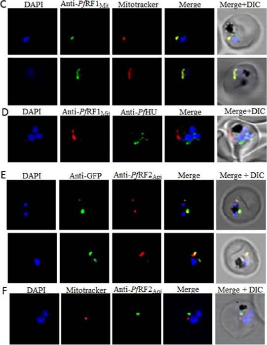
Detection and subcellular localization of PfRF1Mit and PfRF2Api. (C-F) Immunofluorescence localization of the RFs in P. falciparum infected erythrocytes by confocal microscopy. PfRF1Mit co-localizes with Mitotracker Red (C), with no overlap seen with the apicoplast marker PfHU (D). PfRF2Api colocalizes with apicoplast-targeted GFP in the P. falciparum D10 ACPleader-GFP line. (E); no overlap of the PfRF2Api signal is seen with Mitotracker Red (F). Nuclear DNA is stained with DAPI.Vaishya S, Kumar V, Gupta A, Siddiqi MI, Habib S. Polypeptide release factors and stop codon recognition in the apicoplast and mitochondrion of Plasmodium falciparum. Mol Microbiol. 2016 Mar 4. [Epub ahead of print]
See original on MMPMore information
| PlasmoDB | PBANKA_0416700 |
| GeneDB | PBANKA_0416700 |
| Malaria Metabolic Pathways | Localisation images Pathways mapped to |
| Previous ID(s) | PB000792.02.0, PBANKA_041670 |
| Orthologs | PCHAS_0417600 , PF3D7_0904700 , PKNH_0702400 , PVP01_0703100 , PVX_098680 , PY17X_0419500 |
| Google Scholar | Search for all mentions of this gene |