PBANKA_0416000 high molecular weight rhoptry protein 3, putative (RhopH3)
Disruptability [+]
| Species | Disruptability | Reference | Submitter | |
|---|---|---|---|---|
| P. berghei ANKA |
Refractory |
PlasmoGEM (Barseq) | PlasmoGEM | |
| P. falciparum 3D7 |
Refractory |
10878256 | Theo Sanderson, Wellcome Trust Sanger Institute | |
| P. falciparum 3D7 |
Refractory |
28221136 CRISPR-mediated |
Theo Sanderson, Wellcome Trust Sanger Institute | |
| P. falciparum 3D7 |
Refractory |
USF piggyBac screen (Insert. mut.) | USF PiggyBac Screen | |
Mutant phenotypes [+]
| Species | Stage | Phenotype | Reference | Submitter |
|---|---|---|---|---|
| P. falciparum 3D7 | Asexual |
Invasion defect |
28252384 Conditional truncation diminished invasion, those parasites which did invade had deficient nutrient import |
Theo Sanderson, Wellcome Trust Sanger Institute |
| P. falciparum 3D7 | Asexual |
Invasion defect |
28221136 (Knock down)
glmS knockdown yielded dose-dependent invasion inhibition. Normal egress. CLAG3 was also lost upon the knockdown. |
Theo Sanderson, Wellcome Trust Sanger Institute |
Imaging data (from Malaria Metabolic Pathways)
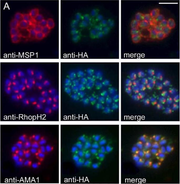
PfSUB2 Is a Microneme Protein. (A) IFA images of schizonts of PfSUB2HA clone 2D dual-labelled with mAbs X509 (anti-MSP1), 61.3 (anti-RhopH2), or 4G2 (anti-AMA1), plus in each case mAb 3F10 (anti-HA) The anti-HA signal co-localised only with the anti-AMA1 signal. Identical results were obtained with the uncloned transgenic PfSUB2HA line, and/or when anti-RAP2 mAb H5 was used as the rhoptry marker instead of mAb 61.3 (not shown). Parasite nuclei are stained throughout with DAPI (blue). Scale bar represents 2 lm.Harris PK, Yeoh S, Dluzewski AR, O'Donnell RA, Withers-Martinez C, Hackett F, Bannister LH, Mitchell GH, Blackman MJ. Molecular identification of a malaria merozoite surface sheddase. PLoS Pathog. 2005 1(3):241-51.
See original on MMP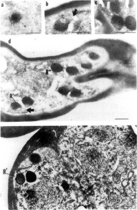
Immunoelectronmicroscopy of rhoptries from schizont-infected erythrocytes at different stages. Cells were fixed in 0.05% glutaraldehyde in 0.1 M cacodylate buffer for 15 min on ice and processed for immunostaining with mAblB9 and protein A-gold. (a) Rhoptry present in 4-nucleus schizont; (b) rhoptry present in 8-nucleus schizont; (c) rhoptry present in 16-nucleus schizont; (d) 8-nucleus schizont showing immature rhoptry, R' and mature rhoptry, R, arrow denotes reticular network; (e) 8-nucleus schizont fixed in 2% glutaraldehyde in 0.1 M cacodylate buffer to preserve morphology and stained with uranyl acetate. Bars, 250 nm.Jaikaria NS, Rozario C, Ridley RG, Perkins ME. Biogenesis of rhoptry organelles in Plasmodium falciparum. Mol Biochem Parasitol. 1993 57(2):269-79. Copyright Elsevier
See original on MMP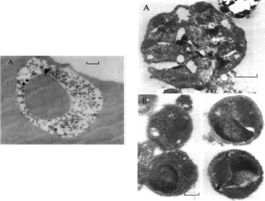
Immunoelectronmicroscopic studies were performed to confirm that the punctate appearance seen by indirect immunofluorescence was indeed due to localization of the target antigen in the rhoptries. Right: FC27 parasites obtained from asynchronous culture were sectioned and incubated with affinity purified human anti-Ag44 antibodies and subsequently with protein-A gold. Gold particles were localized within paired organelles at the apex of both immature merozoites within segmented schizonts (A) and free merozoites (B). These organelles had the typical appearance of rhoptries. Bottom: When ring-stage parasites were labelled with anti-Ag44 antibodies, label was found around the developing ring and scattered through the erythrocyte cytoplasm.Coppel RL, Bianco AE, Culvenor JG, Crewther PE, Brown GV, Anders RF, Kemp DJ. A cDNA clone expressing a rhoptry protein of Plasmodium falciparum. Mol Biochem Parasitol. 1987 25:73-81. Copyright Elsevier 2010.
See original on MMP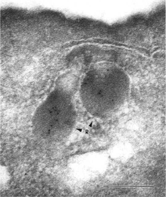
Immunoelectron microscopy on mature schizont-stage parasites was carried out on samples of parasite culture fixed with 0.05% (v/v) glutaraldehyde and embedded in LR White resin, medium grade. The sections were etched and immunolabeled. The immunolabeled sections were contrasted using lead citrate and uranyl acetate.Cooper JA, Ingram LT, Bushell GR, Fardoulys CA, Stenzel D, Schofield L, Saul AJ. The 140/130/105 kilodalton protein complex in the rhoptries of Plasmodium falciparum consists of discrete polypeptides. Mol Biochem Parasitol. 1988 29:251-60. Copyright Elsevier 2010.
See original on MMP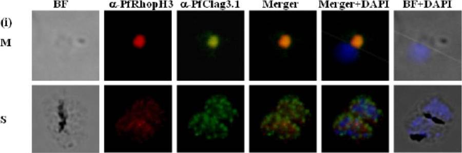
Immunofluorescence assay to show localization of PfRhopH3-C in rhoptries. (i) Co-localization of PfRhopH3 and PfClag3.1 using their respective antibodies at schizont (S) and free merozoites (M) stages. Staining with preimmune sera and anti-GST antibodies respectively were used as controls. The parasite nuclei were stained with DAPI (blue). PfRhopH3 and PfClag3.1 were localized at the apical tip.Ranjan R, Chugh M, Kumar S, Singh S, Kanodia S, Hossain MJ, Korde R, Grover A, Dhawan S, Chauhan VS, Reddy VS, Mohmmed A, Malhotra P. Proteome analysis Rrveals a large merozoite surface protein-1 associated complex on the Plasmodium falciparum merozoite surface. J Proteome Res. 2010 10(2):680-91 Copyrihgt American Chemical Society 2011.
See original on MMP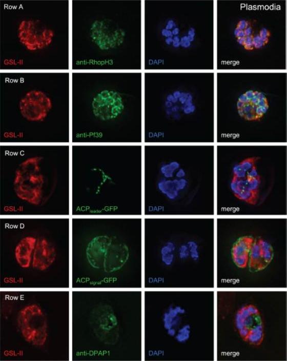
Deconvolving micrographs of Plasmodium-infected RBCs show that GSL-II, which binds to Plasmodium N-glycans, colocalizes with rhoptries (row A) and the ER (row B) but does not colocalize with apicoplasts (row C), the parasitophorous vacuole (row D), or the food vacuole (row E). In each case GSL-II is labeled red with Alexafluor 594, while antibodies labeled green with Alexafluor 488 are against RhopH3 (rhoptries), Pf39 (ER), and food vacuole (DPAP1). Alternatively, GFP is targeted to apicoplasts (ACPleader-GFP) and the parasitophorous vacuole (ACPsignal-GFP). Control experiments with anti-GFP antibodies show that both apicoplasts and parasitophorous vacuoles are accessible to exogenous probes. The nature of the glycosylated proteins is not knownBushkin GG, Ratner DM, Cui J, Banerjee S, Duraisingh MT, Jennings CV, Dvorin JD, Gubbels MJ, Robertson SD, Steffen M, O'Keefe BR, Robbins PW, Samuelson J. Suggestive evidence for Darwinian Selection against asparagine-linked glycans of Plasmodium falciparum and Toxoplasma gondii. Eukaryot Cell. 2010 9:228-41.
See original on MMP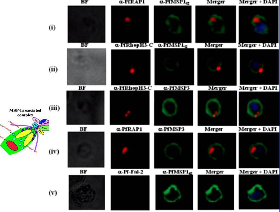
Localization of proteins of PfMSP-1 associated complex by immunofluorescence assay. Unpermeabilized P. falciparum merozoites were coimmunostained with anti-PfRAP-1 and anti-PfMSP-142 antibodies (i), with anti-PfRhopH3-C and anti-PfMSP-142 antibodies (ii), with anti-PfRhopH3-C and anti-PfMSP-3 antibodies (iii), anti-PfRAP-1 and anti-PfMSP-3 antibodies (iv), and anti-Pffalcipain-2 and anti-PfMSP-142 antibodies (v). PfMSP-1 and PfMSP-3 staining was detected on the entire surface of merozoites, while PfRhopH3 and PfRAP-1 staining localized at the apical end of merozoites. Co-localization was observed between PfMSP-1 and PfRAP-1, PfMSP-1 and PfRhopH3, PfMSP-3 and PfRhopH3 as well PfMSP-1 and PfMSP-3 staining was detected on the entire surface of merozoites, while PfRhopH3 and PfRAP-1 staining localized at the apical end of merozoites. Co-localization was observed between PfMSP-1 and PfRAP-1, PfMSP-1 and PfRhopH3, PfMSP-3 and PfRhopH3 as well antibody and the specific location of PfRhopH3 in rhoptries. Ranjan R, Chugh M, Kumar S, Singh S, Kanodia S, Hossain MJ, Korde R, Grover A, Dhawan S, Chauhan VS, Reddy VS, Mohmmed A, Malhotra P. Proteome analysis Rrveals a large merozoite surface protein-1 associated complex on the Plasmodium falciparum merozoite surface. J Proteome Res. 2010 10:680-91. Copyright American Chemical Society
See original on MMP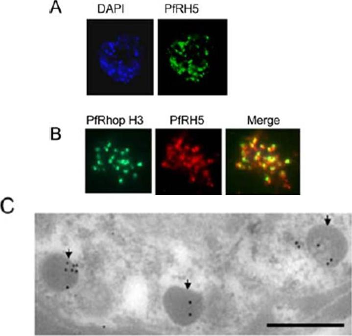
Apical localization of PfRH5 in the merozoite. A Mature Dd2 schizonts were labeled with rabbit anti-PfRH5 IgG and counterstained with FITC-labeled anti-rabbit IgG (green) using immunofluorescence microscopy. B Colocalization studies: Mature Dd2 schizonts were double-labeled with anti-PfRH5 IgG (red) and anti-RhopH3 (green) monoclonal antibody. The merged staining (yellow) by both antibodies indicates their similar location within the parasite. C Immunoelectron microscopy confirming the localization of PfRH5 in the rhoptries within the developing merozoites in the Dd2 schizont. Parasites were fixed with 1% paraformaldehyde and 0.1% glutaraldehyde. Staining was detected with rabbit anti-RH5 and antirabbit gold (10 nm). Scale bar represents 500 nm.Rodriguez M, Lustigman S, Montero E, Oksov Y, Lobo CA. PfRH5: a novel reticulocyte-binding family homolog of plasmodium falciparum that binds to the erythrocyte, and an investigation of its receptor. PLoS One. 2008 3(10):e3300.
See original on MMP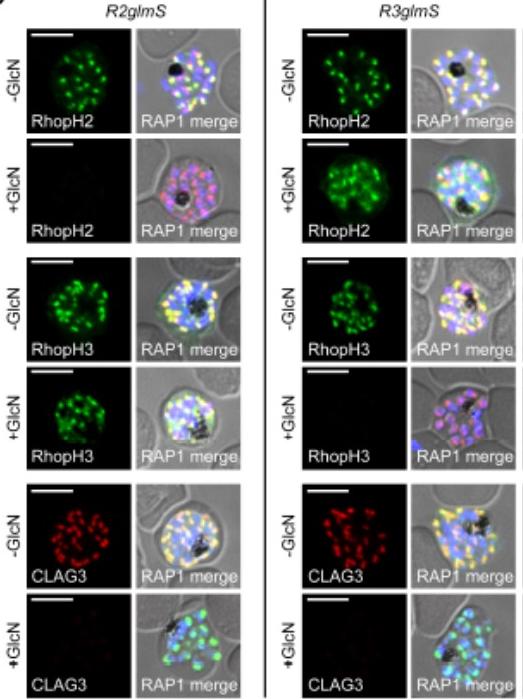
IFA of indicated proteins in R2glmS (RopH2 knockdown) and R3glmS (RopH3 knockdown) at schizont stage; colocalization with RAP1 and apical puncta in daughter merozoites indicates rhoptry trafficking. Scale bars, 5 μm. GlcN treatment of either R2glmS or R3glmS abolished not only the targeted gene product but also CLAG3, a third better-characterized member of the RhopH complex. Indirect immunofluorescence microscopy confirmed this surprising finding and provided additional insights. While RhopH2 was still detected in the R3glmS knockdown, it did not fully colocalize with RAP1 but instead had a more diffuse distribution; this suggests RhopH2 may be retained in the parasite endoplasmic reticulum if it fails to interact with RhopH3. In contrast, RhopH3 appeared to traffic normally inGlcN-treated R2glmS parasites, as indicated by apical colocalization with RAP1 inmerozoites. Thus, CLAG3, RhopH2 and RhopH3 assemble into a complex either during or immediately after translation.Ito D, Schureck MA, Desai SA. An essential dual-function complex mediates erythrocyte invasion and channel-mediated nutrient uptake in malaria parasites. Elife. 2017 Feb 21;6.
See original on MMP
RhopH proteins are deposited in the PV and exported via PTEX. IFA of 13F10 showing export of RhopH proteins after growth with or without TMP for 48 h. Colocalization with exported HRP2 or SBP1, as indicated. Scale bars, 5 μm. Export of RhopH2 and RhopH3 were both fully blocked in trophozoite-stage parasites grown for 48 h without TMP. Colocalization with HRP2, a parasite protein carrying the canonical PEXEL export signal showed both proteins trapped in the vacuole upon PTEX knockdown. CLAG3 export requires PTEX activity as TMP removal prevented its movement out of the PV (bottom row); colocalization with SBP1, another PTEX substrate.Ito D, Schureck MA, Desai SA. An essential dual-function complex mediates erythrocyte invasion and channel-mediated nutrient uptake in malaria parasites. Elife. 2017 Feb 21;6.
See original on MMP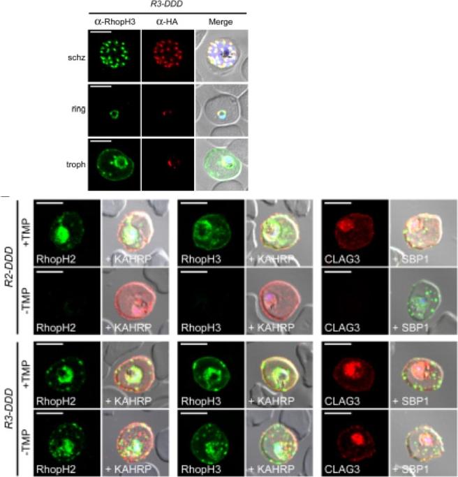
(D) IFA of R3-DDD (conditional RopH3 mutant) probed with indicated antibodies at each parasite stage. Note that both antibodies detect protein in schizont rhoptries, but that only the anti-RhopH antibody detects export to the host membrane in trophozoites. Scale bars, 5 μm. (E) Trophozoite-stage IFAs of indicated parasites cultivated with and without TMP for 48 h, showing that knockdown abolishes each member of the complex in R2-DDD (conditional RopH2 mutant) but not R3-DDD. Co-localization is shown with exported parasite proteins KAHRP or SBP1 (red or green in merge panels, respectively). Scale bars, 5 μm. The RhopH complex appears to be secreted into the PV (upper panel), suggesting that its member proteins must cross the bounding PVM to enter host cytosol and eventually reach the erythrocyte surface. While TMP removal rendered all three RhopH proteins undetectable in the R2-DDD parasite, these proteins and their transfer to trophozoites were preserved inR3-DDD.Ito D, Schureck MA, Desai SA. An essential dual-function complex mediates erythrocyte invasion and channel-mediated nutrient uptake in malaria parasites. Elife. 2017 Feb 21;6. PMID: 28221136.
See original on MMP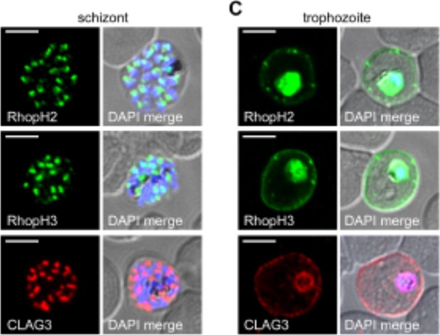
RhopH2 and RhopH3 are membrane-associated proteins. IFA of indicated proteins in R2glmS (RopH2 knockdown) and R3glmS (RopH3 knockdown) at the trophozoite stage. IFA of mature schizont-infected cells with indicated antibodies. Punctate labeling indicates that these proteins localize to rhoptry organelles in daughter merozoites. Scale bar, Scale bars, 5 μm. IFA of trophozoite-infected cells, showing that each protein localizes to the host membrane and to the vacuole surrounding the intracellular parasite. Scale bar, 5 μmImmunofluorescence microscopy confirmed packaging into rhoptries within schizont-infected cells (B). Imaging of maturing trophozoite-stage parasites also showed that these proteins traffic to the host membrane (C). Because each gene is transcribed and translated only within schizonts, the protein found in these maturing trophozoites reflects transfer from the merozoite during invasion.Ito D, Schureck MA, Desai SA. An essential dual-function complex mediates erythrocyte invasion and channel-mediated nutrient uptake in malaria parasites. Elife. 2017 Feb 21;6.
See original on MMP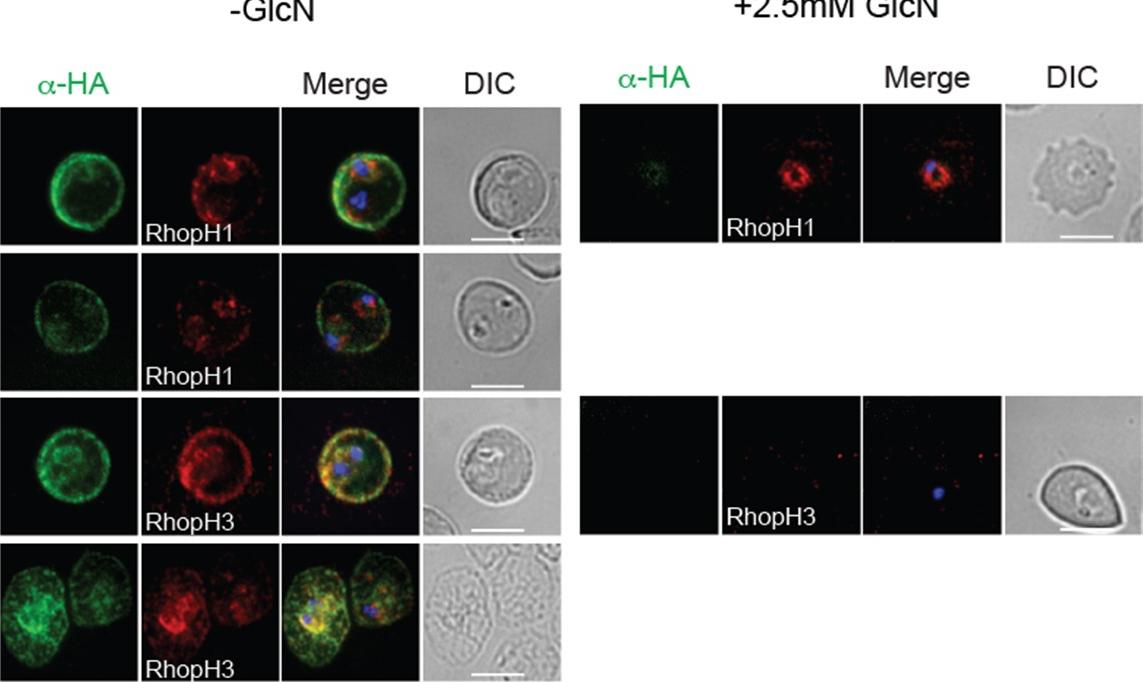
Trafficking of apical proteins is not affected by RAP1 knockdown IFA of schizont stage parasites showing that trafficking of rhoptry (RAMA, RHOPH3, RON6) and micronemal (AMA1) proteins is not affected when RAP1 or RAP2 expression is depleted from parasites with anhydrotetracycline )Atc(. No apparent difference in the electron density or morphology of the rhoptries was observed (Left panel), suggesting that RAP2 is not essential for rhoptry formation in Plasmodium spp. Moreover, knockdown of either RAP1 or RAP2 expression did not affect parasite maturation and replication, as there was no significant difference observed in merozoite number or schizont formation compared to parasites grown in the absence of ATc (right panel).Ghosh S, Kennedy K, Sanders P, Matthews K, Ralph SA, Counihan NA, de Koning-Ward TF. The Plasmodium rhoptry associated protein complex is important for parasitophorous vacuole membrane structure and intraerythrocytic parasite growth. Cell Microbiol. 2017.
See original on MMP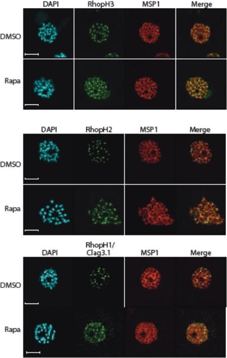
Truncation of RhopH3 leads to mistrafficking of components of the RhopH complex. IFA of mature schizonts of control (DMSO) and rapamycin-treated rhopH3-loxP parasites, probed with MSP1-specific antibodies (either mAb 89.1 or rabbit polyclonal anti-MSP1 antibodies; red) and antibodies to the three indicated RhopH components (green). Mis-localisation of the RhopH proteins was observed in all cases, and in the case of RhopH3 the protein often appeared to reside external to the plasma membrane of intracellular merozoites. Parasite nuclei were visualized by staining with DAPI. Note that, for clarity, the merge panels do not include the DAPI signal. Scale bar, 5 μm.Sherling ES, Knuepfer E, Brzostowski JA, Miller LH, Blackman MJ, van Ooij C. The Plasmodium falciparum rhoptry protein RhopH3 plays essential roles in host cell invasion and nutrient uptake. Elife. 2017 6. pii: e23239.
See original on MMP
Truncation of RhopH3 leads to mistrafficking of components of the RhopH complex and loss of complex formation. A) IFA showing colocalization of RhopH3, RhopH2 and RhopH1/Clag3.1 with the rhoptry marker RAP2 in schizonts of control (DMSO) rhopH3-loxP parasites but loss of colocalization following rapamycin (Rapa) treatment. Parasite nuclei were visualized by staining with 4,6-diamidino-2-phenylindole (DAPI). Scale bar, 5 μm. Immunofluorescence analysis (IFA) showed that, as expected, RhopH3 colocalized with the rhoptry marker RAP2 in mature schizonts of control rhopH3-loxP parasites. However, in rapamycin-treated (RhopH3Δ4-219 6) parasites, this colocalization was lost, although RAP2 was still detected in a punctate, apically-disposed pattern typical of rhoptries B) Colocalization of the members of the RhopH complex. RhopH3, RhopH2 and Rhop 1/Clag3.1 colocalize in rhopH3-loxP parasites treated with DMSO, but this colocalization is lost in parasites treated with rapamycin. Scale bar, 2 μm. Neither RhopH2 nor RhopH1/Clag3.1 colocalized with 233 RhopH3Δ4-6 in the mutant parasites. The RhopH2 and RhopH1/Clag3.1 signals were also distinct in the mutant parasites, although in this case some limited colocalization of these proteins was apparent (bottom images).Sherling ES, Knuepfer E, Brzostowski JA, Miller LH, Blackman MJ, van Ooij C. The Plasmodium falciparum rhoptry protein RhopH3 plays essential roles in host cell invasion and nutrient uptake. Elife. 2017 6. pii: e23239.
See original on MMP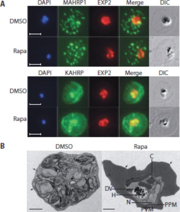
Loss of the RhopH complex does not ablate parasite protein export. Cycle 2 (72 h post rapamycin treatment) DMSO-treated and rapamycintreated rhopH3-loxP clone 5F5 trophozoite-stage parasites were probed withantibodies against the parasitophorous vacuole membrane marker EXP2 to delineate the parasite in the infected erythrocyte, as well as antibodiesspecific for either the Maurer’s cleft marker MAHRP1 (top panels) or the export marker KAHRP (bottom panels). Scale bar, 5 μm. B) Transmission electron micrograph showing a comparison between cycle 2 parasites ofDMSO-treated or rapamycin-treated rhopH3-loxP clone 5F5 parasites ~ 92 h following rapamycin treatment. The developmental block in the RhopH3Δ4-6 parasite is clearly evident, as is the presence of knobs (arrowed) on thesurface of the erythrocyte in both cases. Components of the mutant parasite labelled are the digestive vacuole (DV), haemozoin (H), nucleus (N), parasitophorous vacuole membrane (PVM), cytostomes (C) and parasite plasma membrane (PPM). The mutant parasites displayed no obvious ultrastructural differences from wild-type trophozoites at a similar developmental stage (not shown). Scale bar, 1 μm.Sherling ES, Knuepfer E, Brzostowski JA, Miller LH, Blackman MJ, van Ooij C. The Plasmodium falciparum rhoptry protein RhopH3 plays essential roles in host cell invasion and nutrient uptake. Elife. 2017 6. pii: e23239. PMID: 28252384
See original on MMP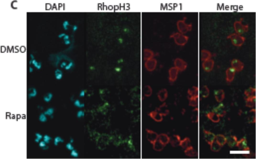
Truncation of RhopH3 leads to mistrafficking of components of theRhopH complex and loss of complex formation. Mislocalisation and reduced levels of RhopH3 in naturally released free merozoites of rhopH3-loxP parasites treated with rapamycin. Samples were probed with a monoclonal antibody to the merozoite surface marker MSP1 as well as anti-RhopH3 antibodies. Scale bar, 2 μm. IFA of naturally released free merozoites showed that the truncated RhopH3Δ4-6 was often largely undetectable in merozoites of the mutant Parasites.Sherling ES, Knuepfer E, Brzostowski JA, Miller LH, Blackman MJ, van Ooij C. The Plasmodium falciparum rhoptry protein RhopH3 plays essential roles in host cell invasion and nutrient uptake. Elife. 2017 6. pii: e23239.
See original on MMP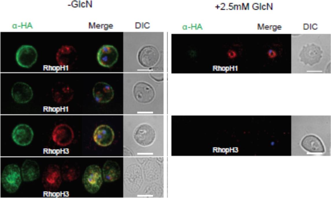
Localization of RhopH1/clag3 and RhopH3 in infected erythrocytes when RhopH2 expression is knocked down. Representative immunofluorescence analysis of erythrocytes infected with RhopH2-HAglmS parasites grown in 0 mM or 2.5 mM GlcN. Cells fixed with acetone/methanol and labelled with anti-HA antibody to detect RhopH2 IFAs and other antibodies, as indicated. the localization of RhopH3 and to a lesser extent RhopH1/clag3 was affected when RhopH2 expression was knocked down.Counihan N, Chisholm SA, Bullen HE, Srivastava A, Sanders PR, Jonsdottir TK, Weiss GE, Ghosh S, Crabb BS, Creek DJ, Gilson PR, de Koning-Ward TF. Plasmodium falciparum parasites deploy RhopH2 into the host erythrocyte to obtain nutrients, grow and replicate. Elife. 2017 Mar 2;6. pii: e23217
See original on MMP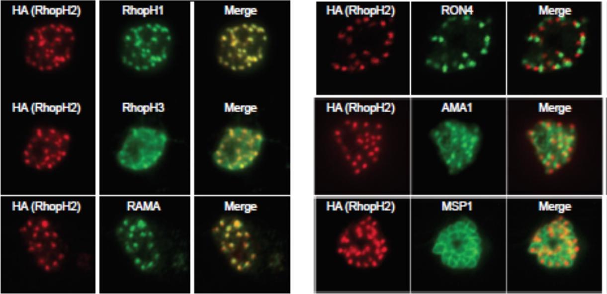
Generation of transgenic parasites in which RhopH2 is epitope-tagged. Immunofluorescence analysis (IFA) on schizonts fixed with acetone/methanol and labelled with anti-HA antibody to detect RhopH2 and other antibodies, as indicated. Immunofluorescence analysis (IFA) confirmed RhopH2-HA localized to the rhoptry and co-localized with other rhoptry bulb proteins, RhopH1, RhopH3 and RAMA but not with the rhoptry neck protein, RON4, the micronemal marker, AMA-1 or the plasma membrane protein MSP1.Counihan N, Chisholm SA, Bullen HE, Srivastava A, Sanders PR, Jonsdottir TK, Weiss GE, Ghosh S, Crabb BS, Creek DJ, Gilson PR, de Koning-Ward TF. Plasmodium falciparum parasites deploy RhopH2 into the host erythrocyte to obtain nutrients, grow and replicate. Elife. 2017 Mar 2;6. pii: e23217
See original on MMP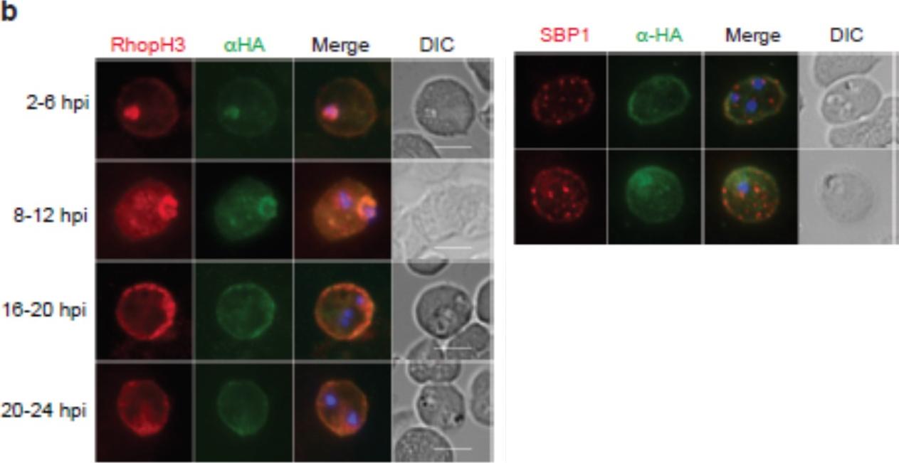
Expression, localisation and solubility profile of P. falciparum RhopH2. Immunofluorescence analysis (IFA) on erythrocytes infected with PfRhopH2-HAglmS and fixed with acetone/methanol. RhopH2 is labeled with the anti-HA antibody. The bars represent 5 μm. (c) IFA on erythrocytes infected with PfRhopH2-HAglmS, fixed with acetone/methanol and probed with anti-HA (for RhopH2) and antibodies to the Maurer’s cleft protein SBP1 show that RhopH2 and SBP1 do not co-localize.Counihan N, Chisholm SA, Bullen HE, Srivastava A, Sanders PR, Jonsdottir TK, Weiss GE, Ghosh S, Crabb BS, Creek DJ, Gilson PR, de Koning-Ward TF. Plasmodium falciparum parasites deploy RhopH2 into the host erythrocyte to obtain nutrients, grow and replicate. Elife. 2017 Mar 2;6. pii: e23217.
See original on MMPMore information
| PlasmoDB | PBANKA_0416000 |
| GeneDB | PBANKA_0416000 |
| Malaria Metabolic Pathways | Localisation images Pathways mapped to |
| Previous ID(s) | PB000377.03.0, PB001160.02.0, PB300291.00.0, PBANKA_041600 |
| Orthologs | PCHAS_0416900 , PF3D7_0905400 , PKNH_0703100 , PVP01_0703800 , PVX_098712 , PY17X_0418800 |
| Google Scholar | Search for all mentions of this gene |