PBANKA_0211300 cysteine desulfurase, putative (NFS)
Mutant phenotypes [+]
None reported yet. Please press the '+' button above to add one.Imaging data (from Malaria Metabolic Pathways)
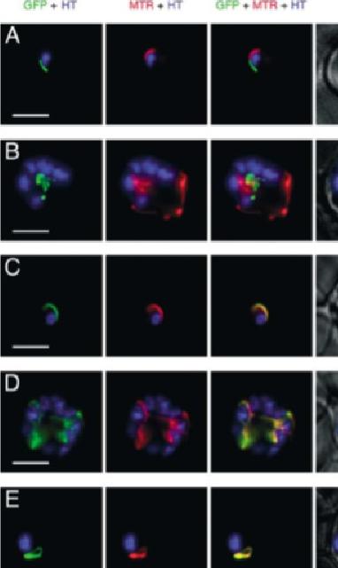
GFP expressed in transfectants of P. falciparum. RBC infected by the transfectant with pSSPF2/PfACP-GFP (A and B), pSSPF2/PfHsp60-GFP (C and D) or pSSPF2/PfIscS-GFP (E and F) were incubated in RPMI 1640 medium containing Hoechst 33342 (HT: blue) at 1 mg/ml and MitoTracker Red CM-H2XRos (MTR: red) at 100nM (A, C, E) or 10nM (B, D, F) to stain the nucleus and mitochondrion, respectively. Localization of GFP (green) in the transfectant parasites in the trophozoite (A, C, E) or schizont (B, D, F) stage was monitored by fluorescence microscopy using a DeltaVison microscope system (Applied Precision). +phase: GFP + MTR + HT superimposed on a phase contrast image. Scale bar: 5 mm. Both Hsp60 and IscS target to the mitochondrion.Sato S, Rangachari K, Wilson RJ. Targeting GFP to the malarial mitochondrion. Mol Biochem Parasitol. 2003 130:155-8 Copyright Elsevier
See original on MMP
Subcellular localization of IscS and Isd11 to the mitochondrion of P. falciparum. A) Epifluorescent images of live P. falciparum erythrocytic-stage parasites expressing GFP fused to full-length IscS (IscSfl-GFP). The parasites were stained with mitotracker to identify mitochondria and DAPI to identify nuclei. Image z-stacks were deconvolved and then presented as a single combined image. Scale bar = 2 mm. B) Epifluorescent images similar to those in (A) of parasites expressing GFP fused to full length Isd11 (Isd11fl-GFP). IscS and Isd11 co-localize with mitotracker in all erythrocytic stages: late ring (top panels), late trophozoite or early schizont (middle panels), and schizont (lower panels) stage parasites. Lowest panel) Subcellular localization of the IscS leader peptide to the mitochondrion of P. falciparum. Epifluorescent images of live P. falciparum erythrocytic-stage parasites expressing GFP fused to the leader peptide of IscS (amino acids 1–35). The parasites were stained with mitotracker to identify mitochondria and DAPI to identify nuclei. Image z-stacks were deconvolved and then presented as a single combined image. Scale bar = 2 µm.Gisselberg JE, Dellibovi-Ragheb TA, Matthews KA, Bosch G, Prigge ST. The Suf Iron-Sulfur Cluster Synthesis Pathway Is Required for Apicoplast Maintenance in Malaria Parasites. PLoS Pathog. 2013 9(9):e1003655
See original on MMP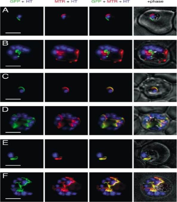
GFP expressed in transfectants of P. falciparum. RBC infected by the transfectant with pSSPF2/PfACP-GFP (A and B), pSSPF2/PfHsp60-GFP (C and D) or pSSPF2/PfIscS-GFP (E and F) were incubated in RPMI 1640 medium containing Hoechst 33342 (HT: blue) at 1 mg/ml and MitoTracker Red CM-H2XRos (MTR: red) at 100nM (A, C, E) or 10nM (B, D, F) to stain the nucleus and mitochondrion, respectively. Localization of GFP (green) in the transfectant parasites in the trophozoite (A, C, E) or schizont (B, D, F) stage was monitored by fluorescence microscopy using a DeltaVison microscope system (Applied Precision). +phase: GFP + MTR + HT superimposed on a phase contrast image. Scale bar: 5 mm. Both Hsp60 and IscS target to the mitochondrion.Sato S, Rangachari K, Wilson RJ. Targeting GFP to the malarial mitochondrion. Mol Biochem Parasitol. 2003 130:155-8.
See original on MMPMore information
| PlasmoDB | PBANKA_0211300 |
| GeneDB | PBANKA_0211300 |
| Malaria Metabolic Pathways | Localisation images Pathways mapped to |
| Previous ID(s) | PB000702.00.0, PBANKA_021130 |
| Orthologs | PCHAS_0209700 , PF3D7_0727200 , PKNH_0211800 , PVP01_0212900 , PVX_081665 , PY17X_0212700 |
| Google Scholar | Search for all mentions of this gene |